A Basic Demonstration Of Optical Cloaking (by UniversityRochester)
A Basic Demonstration of Optical Cloaking (by UniversityRochester)
More Posts from Fuadalanazi and Others

يالله 💙
BugsFeed: 7 bad ass organisms that can survive intracellularly in immune cells
1. Mycobacterium tuberculosis - Stops fusion!

Mycobacterium tuberculosis utilizes macrophages for its replication! (It uses the usual killer to expand it’s army :O ) How does tuberculosis bacilli survive in macrophages? M. tuberculosis has evolved a number of very effective survival strategies - It inhibits phagosome-lysosome fusion and inhibits phagosome acidification ensuring it’s survival inside the macrophage.
2. Brucella - Has chains, like Bruce Lee.

Brucella has a LPS O-chain. It ensures the Brucella containing vacuole (BCV) avoids fusion with lysosomes, prevents the deposition of complement at the bacterial surface and forms stable large clusters with MHC-II named macrodomians in the cell surface, interfering with MHC-II presentation of peptides to specific CD4+ T cells. Woah.
3. Listeria - It gets internalized in a vacuole and then runs away.

The pore-forming protein listeriolysin O mediates escape from host vacuoles. Once in the cytosol, the L. monocytogenes mediates efficient actin-based motility, thereby propelling the bacteria into neighboring cells. The cytosol is a favorable environment for listeria’s growth.
4. Mycobacterium leprae - Cholesterol and TACO!

Mycobacterium leprae is able to induce lipid droplet formation in infected macrophages. Cholesterol mediates the recruitment of TACO from the plasma membrane to the phagosome. TACO, also termed as coronin-1A (CORO1A), is a coat protein that prevents phagosome-lysosome fusion and thus degradation of mycobacteria in lysosomes. The entering of mycobacteria at cholesterol-rich domains of the plasma membrane and their subsequent uptake in TACO-coated phagosomes promotes intracellular survival.
5. Coxiella brunetti - The indestrucible

This hardy, obligate intracellular pathogen has evolved to not only survive, but to thrive, in the harshest of intracellular compartments: the phagolysosome. Following internalization, the nascent Coxiella phagosome ultimately develops into a large and spacious parasitophorous vacuole (PV) that acquires lysosomal characteristics such as acidic pH, acid hydrolases and cationic peptides, defences designed to rid the host of intruders.
6. Salmonella - TTSS

Salmonella have a specialized secretion system, termed the type III secretion system (TTSS), as well as proteins secreted by this system, are encoded in Salmonella pathogenicity island 1 (SPI1). TTSS are used by bacterial pathogens to inhibit their phagocytosis, induce eukaryotic cell death, and alter the host cell cytoskeleton. Salmonella species have at least one other TTSS encoded on SPI2 that appears to be involved in intracellular survival.
7. Human Immunodeficiency Virus - Tries to not attract attention

After infecting cells, HIV survives. Ever wondered why? It’s because the HIV protein, Nef plays a role in downregulating the expression of various proteins needed for recognition by potentially dangerous CD8 T cells. Nef lowers the surface expression of CD4, and several haplotypes of MHC-I by redirecting their transport from the trans-Golgi network. Another gene, Tat, appears to upregulate the expression of Bcl-2 during the early phase of cellular infection, increasing the likelihood that it will receive survival signals.
Many viruses can survive intracellularly, but I’ve included specifically HIV in this list because it survives in immune cells and it is an important virus to know.
For appropriate sources and references, click here.

Frankenstein’s…bacteriophages?
A group of scientists have just published their work in CellPress on synthesising novel phages using common viral structures (Figure 1), to target a range of bacterial hosts. Anyone who frequently reads my posts will be fully aware of what bacteriophages are, however put simply phages are viruses that target bacteria (Figure 1). Currently a lot of research is going into these little guys to utilise them for both therapeutic and diagnostic purposes.

Figure 1. Image depicts a transmission electron micrograph of a phage (left) and a diagrammatic representation of the phage on the (right) showing all the major components that make up the protein coat.
Phages have an elaborate protein coat that surrounds their DNA cargo (contained within the head, Figure 1). They scan their surrounding environment for potential ‘prey’ by using their tail fibres to interact with their cognate receptors. The diversity of receptors is immense and many still go unidentified even for well characterised phages. Once a target is found, the baseplate then irreversibly attaches to the cell wall of the bacteria and the DNA can be transferred into the cytoplasm of the cell.
Unlike current therapeutics, the sheer diversity between phages means that they are highly specific with limited host ranges to the species or even strain level. As a result, for phage therapy to be effective, a cocktail of phages needs to be employed to target a wide range of potential bacterial hosts. To improve the host ranges of phage groups such as this one are trying to synthesis novel structures from existing phages - much like how Frankenstein stitched together a new human body from pre-existing parts (Figure 2).

Figure 2. Simplified image showing how bacterial components can be shuffled between genomes of related phages with different host ranges.
Innovatively, this group used a well characterised yeast-based (Saccharomyces cerevisiae) platform for capturing phage genomes to allow their genetic manipulation (Figure 3). Phage genomes could then be inserted into a yeast artificial chromosome (YAC) then manipulated. Yeast are fairly easy to genetically modify by homologous-recombingation, making this system far easier to employ than other methods. The YAC was then recovered and transformed into the normal host bacteria, allowing the generation of new phage particles. Unlike other methods phage generated via this method were relatively easy to reboot back into active viral particles.

Figure 3. Illustration of the genetic manipulation of phage DNA to generate hybrid phages with altered host ranges.
The group managed to show that by swapping modular components such as tail fibres between phages, new host specificity could be generated. Thus illustrating that gene swapping can overcome strain or species barriers if the need arises. This work will hopefully lead way to further improve phage therapy and decrease the persistent need for the identification of novel phages. Further work needs to be done on increasing the scope of their work, but they have created a framework that will hopefully be able to reboot more synthetic phages.
“Our results show that common phage scaffolds can be re-targeted against new bacterial hosts by engineering single or multiple tail components. This capability enables the the construction of defined phage cocktails that only differ in their host range determinants and can be used to edit the compositions of microbial consortia and/or treat bacterial infections.” - Ando et al, 2015
Sources:
Engineering Modular Viral Scaffolds for Targeted Bacterial Population Editing, 2015. Ando, H., Lemire. S., Pires. D.P., Lu. T.K., Cell Systems , Volume 1 , Issue 3 , 187 - 196


This week’s infographic is a metabolic map showing the chemical reactions that happen inside our body. You can check out the full size version here.
Magnified Photos of Creepy Crawlies

Colorado Potato Beetle magnified 100x. ‘As you might infer from its name, this bug loves potato crops and destroys plenty of them (and sometimes eggplant and tomato crops, too).‘

A schistosome parasite magnified 256x. This parasite ‘can penetrate the skin of people who come into contact with contaminated water. After several weeks, the parasites mature into adult worms, which live and produce eggs in blood vessels.’

‘Looks kind of like a sloth, doesn’t it? A sloth that climbs through your hair (and sometimes your eyebrows and eyelashes) laying eggs. Adult lice are just 2 to 3 mm long; this one has been magnified 200 times.’

This capture of a Bed Bug ‘shows the insect’s mouthparts, which it uses to pierce skin and drink your blood while you sleep.’

The infamous Water Bear, also known as a tardigrade, is much less adorable in real life than it is in many people’s imagination. But no matter how cute you find them, they’re indisputably badasses of nature, showing the ability to survive the vacuum of space.
See more magnified creepy crawlers at Mental_Floss


Cone snails might not seem like deadly predators, especially when you consider how easily a fish could outswim them. However, these snails await the cover of darkness to prey on sleeping fish. They appear to release paralyzing chemicals before using a venomous barb to finally put the fish out of its misery. (Source)


Bacterias & Tx
Hi guys, just wanted to let you know that I did this really fast, so if there’s something wrong let me know! thanks :)

No bacterium is an island.
Many people think of bacteria as tiny Lone Rangers, paddling their flagellar canoes across the desolate petri dish sea. But in “the wild”, bacteria exist as complex, interwoven, constantly competing social communities.
Every scoop of soil is a battlefield of chemical chatter. Species send out molecular messages-in-a-bottle that ride the waves of diffusion to their mates. Some even thread electrical cables between neighboring cells. Now, new research has identified elaborate shared membranes that let single cells swarm as a superorganism …
Check out my latest article for Wired all about a soil bacterium named Myxococcus xanthus. It’s under everyone’s feet right now, and it has developed one of the most elaborate physical webs ever witnessed in bacteria. That’s it up top, devouring a colony of E. coli using its patented rippling wave attack.
It’s a stealth communication network that lets them hunt like a tiny wolfpack. So cool. Plus I got to use a GIF, so double win.
Once you’re done with that, check out this great TED talk from Bonnie Bassler all about how bacteria communicate.
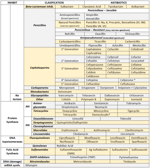
ANTIBIOTICS CHEAT SHEET :)
Also, REMEMBER!!!!
* Sulfonamides compete for albumin with:
Bilirrubin: given in 2°,3°T, high risk or indirect hyperBb and kernicterus in premies
Warfarin: increases toxicity: bleeding
* Beta-lactamase (penicinillase) Suceptible:
Natural Penicillins (G, V, F, K)
Aminopenicillins (Amoxicillin, Ampicillin)
Antipseudomonal Penicillins (Ticarcillin, Piperacillin)
* Beta-lactamase (penicinillase) Resistant:
Oxacillin, Nafcillin, Dicloxacillin
3°G, 4°G Cephalosporins
Carbapenems
Monobactams
Beta-lactamase inhibitors
* Penicillins enhanced with:
Clavulanic acid & Sulbactam (both are suicide inhibitors, they inhibit beta-lactamase)
Aminoglycosides (against enterococcus and psedomonas)
* Aminoglycosides enhanced with Aztreonam
* Penicillins: renal clearance EXCEPT Oxacillin & Nafcillin (bile)
* Cephalosporines: renal clearance EXCEPT Cefoperazone & Cefrtriaxone (bile)
* Both inhibited by Probenecid during tubular secretion.
* 2°G Cephalosporines: none cross BBB except Cefuroxime
* 3°G Cephalosporines: all cross BBB except Cefoperazone bc is highly highly lipid soluble, so is protein bound in plasma, therefore it doesn’t cross BBB.
* Cephalosporines are "LAME“ bc they do not cover this organisms
L isteria monocytogenes
A typicals (Mycoplasma, Chlamydia)
M RSA (except Ceftaroline, 5°G)
E nterococci

* Disulfiram-like effect: Cefotetan & Cefoperazone (mnemonic)
* Cefoperanzone: all the exceptions!!!
All 3°G cephalosporins cross the BBB except Cefoperazone.
All cephalosporins are renal cleared, except Cefoperazone.
Disulfiram-like effect
* Against Pseudomonas:
3°G Cef taz idime (taz taz taz taz)
4°G Cefepime, Cefpirome (not available in the USA)
Antipseudomonal penicillins
Aminoglycosides (synergy with beta-lactams)
Aztreonam (pseudomonal sepsis)
* Covers MRSA: Ceftaroline (rhymes w/ Caroline, Caroline the 5°G Ceph), Vancomycin, Daptomycin, Linezolid, Tigecycline.
* Covers VRSA: Linezolid, Dalfopristin/Quinupristin
* Aminoglycosides: decrease release of ACh in synapse and act as a Neuromuscular blocker, this is why it enhances effects of muscle relaxants.
* DEMECLOCYCLINE: tetracycline that’s not used as an AB, it is used as tx of SIADH to cause Nephrogenic Diabetes Insipidus (inhibits the V2 receptor in collecting ducts)
* Phototoxicity: Q ue S T ion?
Q uinolones
Sulfonamides
T etracyclines

* p450 inhibitors: Cloramphenicol, Macrolides (except Azithromycin), Sulfonamides
* Macrolides SE: Motilin stimulation, QT prolongation, reversible deafness, eosinophilia, cholestatic hepatitis
* Bactericidal: beta-lactams (penicillins, cephalosporins, monobactams, carbapenems), aminoglycosides, fluorquinolones, metronidazole.
* Baceriostatic: tetracyclins, streptogramins, chloramphenicol, lincosamides, oxazolidonones, macrolides, sulfonamides, DHFR inhibitors.
* Pseudomembranous colitis: Ampicillin, Amoxicillin, Clindamycin, Lincomycin.
* QT prolongation: macrolides, sometimes fluoroquinolones