Separation Of A Highly Fluorescent Anthranilic Acid Derivative From The Reaction Mixture.


Separation of a highly fluorescent anthranilic acid derivative from the reaction mixture.
The upper organic layer dissolved almost completely my compound from the reaction mixture and could be separated in one step. A good point was that the compound had a really strong fluorescence and if I placed an UV lamp next to the separation funnel it was easily observed that the water phase contained almost none of the title compound.
More Posts from Contradictiontonature and Others
Phenyllithium or lithobenzene is an organometallic agent with the empirical formula C6H5Li. Crystalline phenyllithium is colorless; however, solutions of phenyllithium are various shades of brown or red depending on the solvent. It is a highly air and moisture sensitive compound, that could be easily decomposed by any protic solvent.

In this case it was a byproduct of a synthesis of an organophosphorous compound and it was only present in a LOW concentration, therefore it was safe to decompose it by simply adding cold water. It’s important to note that it could be dangerous to decompose organometallic compounds by simply adding water. Also, in this case, the highly toxic BENZENE was the product of this reaction, what should be handled with care.


My new favorite: Solvatofluorescence of Nile Red
Solvatochromism is the ability of a chemical substance to change color due to a change in solvent polarity, so it has different color in different solvents.
Also in some cases, the emission and excitation wavelength both shift depending on solvent polarity, so it fluoresces with different color depending on the solvent what it’s dissolved in. This effect is solvatofluorescence.
On the video the highly solvatochromic organic dye, Nile Red was added to different organic solvents and was diluted with another, different polarity organic solvent. As the polarity of the solution changed, the emitted color from the fluorescent dye also varied as seen on the gifs above and as seen on the video:
To help the blog, donate to Labphoto through Patreon: https://www.patreon.com/labphoto

The Portuguese man o’ war delivers a powerful sting to its prey—and sometimes to people—through venom-filled structures on its tentacles. It is not a jellyfish, but rather a colony of different types of zooids (small animals). Jean Louis Coutant engraved the plate for this illustration.
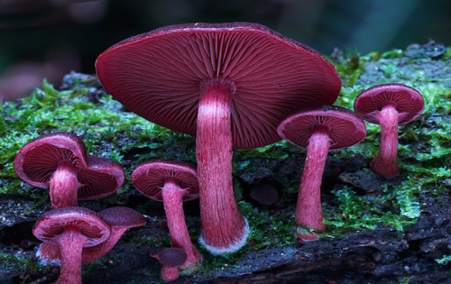
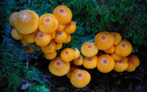
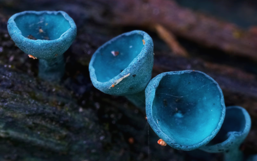
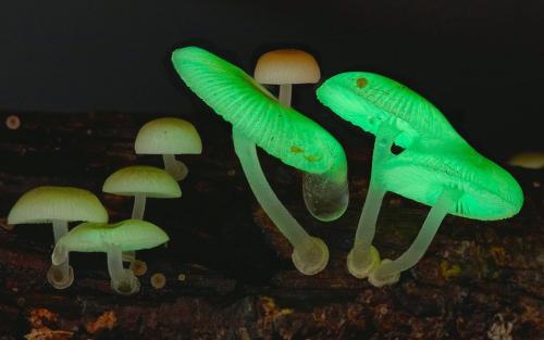

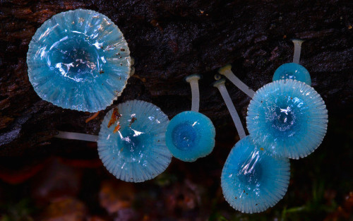
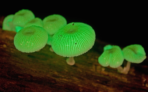
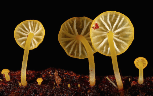
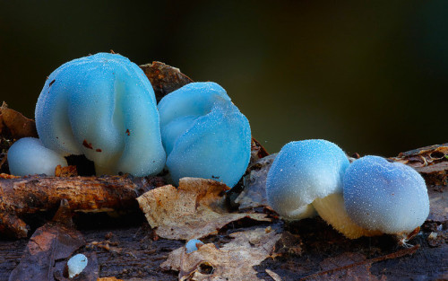
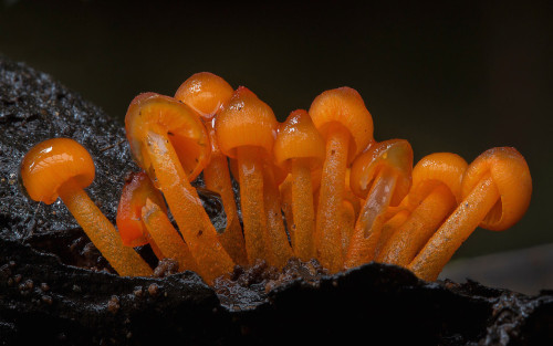
a mushroom rainbow to put the fun in fungi. cause they don’t need psilocybin to be magic. and though some mushrooms are coloured as a toxicity warning to predators, many others are brightly coloured to instead attract potential spore dispersers. see this for more on the bioluminescent mushrooms seen here. (photos)

IN THE MIX
Matti Koivisto, an undergraduate student working in the laboratory of Kari Haajanen at Turku University of Applied Sciences, designs solutions similar to this one to detect the bacteria Escherichia coli. He uses a dye called rhodamine 6G, which has a strong orange color when dissolved in ethanol. In solution, the dye separates into negatively and positively charged ions, the latter of which are largely responsible for the dye’s color. Negatively charged molecules on the outer membranes of E. coli attract the dye’s positive ions. This interaction causes the dye to change color to a pinkish hue, and the level of color change allows Koivisto to gauge how many of the bacteria are present.
Submitted by Matti Koivisto
Do science. Take photos. Make money: Enter our monthly photo contest here for your chance to win!
Related C&EN content:
How dyes in mouse feces help track insects
Using an ink-jet printer to simplify analytical chemistry

As the element that makes up 75 percent of all the mass in the Universe, and more than 90 percent of all the atoms, we’re all pretty well acquainted with hydrogen.
But the simplest and most abundant element in the Universe still has some tricks up its sleeve, because physicists have just created a never-before-seen form of hydrogen - negatively charged hydrogen clusters.
To understand what negatively charged hydrogen clusters are, you first have to wrap your head around their far more common counterparts - positively charged hydrogen clusters.
Positively charged hydrogen clusters are pretty much exactly what they sound like - positively charged clusters of a few or many hydrogen molecules.
Known simply as hydrogen ion clusters, they form at very low temperatures, and can contain as many as 100 individual atoms.
Physicists confirmed the existence of hydrogen ion clusters some 40 years ago, and while a negative counterpart to these clusters boasting large numbers of atoms were theorised, no one could figure out how to create one.
But that didn’t stop a team of physicists led by Michael Renzler from the University of Innsbruck in Austria from giving it a shot.
Continue Reading.

(Image caption: A new technique called magnified analysis of proteome (MAP), developed at MIT, allows researchers to peer at molecules within cells or take a wider view of the long-range connections between neurons. Credit: Courtesy of the researchers)
Imaging the brain at multiple size scales
MIT researchers have developed a new technique for imaging brain tissue at multiple scales, allowing them to peer at molecules within cells or take a wider view of the long-range connections between neurons.
This technique, known as magnified analysis of proteome (MAP), should help scientists in their ongoing efforts to chart the connectivity and functions of neurons in the human brain, says Kwanghun Chung, the Samuel A. Goldblith Assistant Professor in the Departments of Chemical Engineering and Brain and Cognitive Sciences, and a member of MIT’s Institute for Medical Engineering and Science (IMES) and Picower Institute for Learning and Memory.
“We use a chemical process to make the whole brain size-adjustable, while preserving pretty much everything. We preserve the proteome (the collection of proteins found in a biological sample), we preserve nanoscopic details, and we also preserve brain-wide connectivity,” says Chung, the senior author of a paper describing the method in the July 25 issue of Nature Biotechnology.
The researchers also showed that the technique is applicable to other organs such as the heart, lungs, liver, and kidneys.
The paper’s lead authors are postdoc Taeyun Ku, graduate student Justin Swaney, and visiting scholar Jeong-Yoon Park.
Multiscale imaging
The new MAP technique builds on a tissue transformation method known as CLARITY, which Chung developed as a postdoc at Stanford University. CLARITY preserves cells and molecules in brain tissue and makes them transparent so the molecules inside the cell can be imaged in 3-D. In the new study, Chung sought a way to image the brain at multiple scales, within the same tissue sample.
“There is no effective technology that allows you to obtain this multilevel detail, from brain region connectivity all the way down to subcellular details, plus molecular information,” he says.
To achieve that, the researchers developed a method to reversibly expand tissue samples in a way that preserves nearly all of the proteins within the cells. Those proteins can then be labeled with fluorescent molecules and imaged.
The technique relies on flooding the brain tissue with acrylamide polymers, which can form a dense gel. In this case, the gel is 10 times denser than the one used for the CLARITY technique, which gives the sample much more stability. This stability allows the researchers to denature and dissociate the proteins inside the cells without destroying the structural integrity of the tissue sample.
Before denaturing the proteins, the researchers attach them to the gel using formaldehyde, as Chung did in the CLARITY method. Once the proteins are attached and denatured, the gel expands the tissue sample to four or five times its original size.
“It is reversible and you can do it many times,” Chung says. “You can then use off-the-shelf molecular markers like antibodies to label and visualize the distribution of all these preserved biomolecules.”
There are hundreds of thousands of commercially available antibodies that can be used to fluorescently tag specific proteins. In this study, the researchers imaged neuronal structures such as axons and synapses by labeling proteins found in those structures, and they also labeled proteins that allow them to distinguish neurons from glial cells.
“We can use these antibodies to visualize any target structures or molecules,” Chung says. “We can visualize different neuron types and their projections to see their connectivity. We can also visualize signaling molecules or functionally important proteins.”
High resolution
Once the tissue is expanded, the researchers can use any of several common microscopes to obtain images with a resolution as high as 60 nanometers — much better than the usual 200 to 250-nanometer limit of light microscopes, which are constrained by the wavelength of visible light. The researchers also demonstrated that this approach works with relatively large tissue samples, up to 2 millimeters thick.
“This is, as far as I know, the first demonstration of super-resolution proteomic imaging of millimeter-scale samples,” Chung says.
“This is an exciting advance for brain mapping, a technique that reveals the molecular and connectional architecture of the brain with unprecedented detail,” says Sebastian Seung, a professor of computer science at the Princeton Neuroscience Institute, who was not involved in the research.
Currently, efforts to map the connections of the human brain rely on electron microscopy, but Chung and colleagues demonstrated that the higher-resolution MAP imaging technique can trace those connections more accurately.
Chung’s lab is now working on speeding up the imaging and the image processing, which is challenging because there is so much data generated from imaging the expanded tissue samples.
“It’s already easier than other techniques because the process is really simple and you can use off-the-shelf molecular markers, but we are trying to make it even simpler,” Chung says.
Thumpety thump thump thumpety thump thump look at Kinesin go
Myosin, kinesin, and dynein are important proteins governing internal transport. Myosin attached to organelles associates with actin microfilaments to enable the continuous flow of cytoplasm called cytoplasmic streaming.
Kinesins and dynein enable the movement of organelles along microtubules. They attach and move along microtubules. Most kinesins transport organelles from the center towards the periphery of the cell, anterograde transport. Dynein, and a few types of kinesins transport towards the cell center, retrograde transport.
(via thelifeofapremed)


The time will come when diligent research over long periods will bring to light things which now lie hidden. A single lifetime, even though entirely devoted to the sky, would not be enough for the investigation of so vast a subject… And so this knowledge will be unfolded through long successive ages. There will come a time when our descendants will be amazed that we did not know things that are so plain to them… Many discoveries are reserved for ages still to come, when memory of us will have been effaced. Our universe is a sorry little affair unless it has something for every age to investigate… Nature does not reveal her mysteries once and for all.
Seneca
(via scienceisbeauty)
-
 grysi liked this · 1 year ago
 -
 n-hetrocyclic3765 reblogged this · 2 years ago
 -
n-hetrocyclic3765 liked this · 2 years ago
-
 sirelectrodom liked this · 4 years ago
A pharmacist and a little science sideblog. "Knowledge belongs to humanity, and is the torch which illuminates the world." - Louis Pasteur
215 posts