My New Favorite: Solvatofluorescence Of Nile Red


My new favorite: Solvatofluorescence of Nile Red
Solvatochromism is the ability of a chemical substance to change color due to a change in solvent polarity, so it has different color in different solvents.
Also in some cases, the emission and excitation wavelength both shift depending on solvent polarity, so it fluoresces with different color depending on the solvent what it’s dissolved in. This effect is solvatofluorescence.
On the video the highly solvatochromic organic dye, Nile Red was added to different organic solvents and was diluted with another, different polarity organic solvent. As the polarity of the solution changed, the emitted color from the fluorescent dye also varied as seen on the gifs above and as seen on the video:
To help the blog, donate to Labphoto through Patreon: https://www.patreon.com/labphoto
More Posts from Contradictiontonature and Others


The Blue Lava of Kawah Ijen Volcano. The ‘blue lavas’ are a rare phenomenon, only visible on the Kawah Ijen Volcano, in Indonesia. It may look like the volcano is spewing blue lava, but in fact, the shocking blue fire occurs when the volcanic sulphuric gases combust. Emerging from cracks in the volcano’s side, these gases ignite when coming into contact with air. It’s not actual blue lava, but blue flames. (video)
One of the smoothest, most beautiful color changes I’ve ever seen.
The reaction is methoxymethyl deprotection of one of my agonists with concentrated HCl in acetonitrile as my solvent. The color change doesn’t happen in THF!


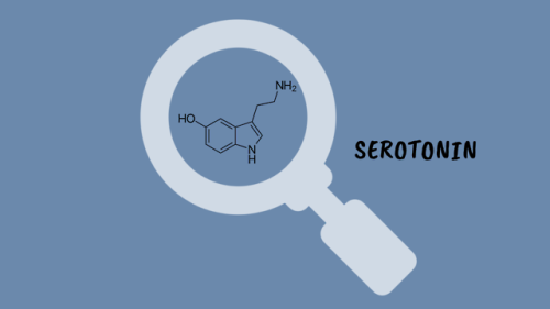
How Do Antidepressants Work? (Video)
Your brain is a network of billions of neurones, all somehow connected to each other. At this very second, millions of impulses are being transmitted through these connections carrying information about what you can see and hear, as well as your emotional state. It’s an incredibly complex system but sometimes things go wrong. Despite extensive research, we are still not certain on the biology that underlies mental illnesses- including depression. However, we have come pretty far in developing effective treatments.

The Hubble Space Telescope captured this picture of the wispy remains of a supernova explosion. The dust cloud in the upper center of the picture is the actual supernova remnant. The dense concentration of stars in the lower left is the outskirts of star cluster NGC 1850. Full resolution picture here. More info here. Credit: NASA, ESA, Y.-H. Chu (Academia Sinica, Taipei)
Checking Cancer At Its Origin..
In a first, the lab led by Leonard Zon at Boston Children’s Hospital has visualised the emergence of the primary melanoma cell in transgenic zebrafish that harbour the human oncogenic BRAFV600E mutation in melanocytes. This cancerous state is characterised in maturing fish by the formation of neural crest progenitors [NCPs], which are the predecessors of melanocytes and are only seen in the embryonic stage of healthy zebrafish.
The Zon lab placed the human mutated oncogene, BRAFV600E (a characteristic of benign human nevi/moles) under the control of a melanocyte-specific promoter and introduced it into the zebrafish. Generations of this transgenic fish were engineered such that they were also deficient in functional p53 (loss of function mutation). They used previous findings that in healthy zebrafish, a gene called crestin is expressed only in the embryonic NCPs and never throughout maturity, but is re-expressed selectively in melanomatous cells during adulthood. crestin was cloned adjacent to a reporter, enhanced green fluorescent protein [EGFP] for live imaging purposes.
The developmental phases of the fish, that were by now triple transgenic (for human BRAFV600E, p53 LOF and crestin:EGFP) were observed by live imaging; ~21 days after fertilisation, the expression of crestin:EGFP localised precisely to the (future) melanoma sites, and the very first triple-transgenic (individual) cells that went on to form larger masses of cells were also observed. To summarise, melanoma formation was observed in three stages: individual fluorescent cells, followed by these cells multiplying to form groups of <50 cells, and lastly these groups forming raised lesions. This consistently held true, with all 30 observed individual cells turning into 30 lesions. These results are illustrated in Figure 1.

Figure 1. In the top left box, a single cell is visualised as it multiplies into a group of melanoma cells (top right). The bottom images show the raised melanoma lesion as observed by the naked eye and by live imaging. The green fluorescence emitted from EGFP indicates that it is localised only to the melanoma (as is crestin expression), that is, it has not metastasised elsewhere.
These pre-cancerous cells were also shown to be self-sustaining and tumourigenic: when fish scales containing the mutant cells were transplanted to another part of the same fish (auto-transplant) or to another fish (allo-transplant) that was also exposed to radiation, the cells proliferated in the new site, as well as penetrated the hypodermis underneath (Figure 2).

Figure 2. The fluorescence indicates a single scale being auto-transplanted elsewhere on the same fish. As the days progress, the patch expands as well, and after day 33, the cells penetrate deeper into the hypodermis and thrive independently, and excising the transplanted scale proves futile.
Role of Transcription Factor sox10
sox10 is a master TF in NCP and its over-expression has been correlated with increased crestin expression, and accordingly, sox10 over expression in the transgenic melanocytes accelerated the melanoma onset. Following the logical train of thought that sox10 promotes melanoma progression, it was then targeted by CRISPR-Cas9 and inactivated in the transgenic cells. This resulted in a delayed onset of melanoma (180 days) compared to the controls (133 days). sox10 is also known to be expressed in most human melanoma cell lines. Moreover, the DNA element that acts as the binding site for Sox10 is also found in a hyper-acetylated [H3K27Ac], super-enhancer state. This is an epigenetic alteration and may prove a useful target in therapy (ex. HAT inhibitors).
Summary
The key finding clears up a hitherto ambiguous association between a reversion to stem/progenitor cell-like status and cancer: it indicates that the apparent devolution of a specialised cell to a primitive cellular state is not a consequence of cancer progression, but that it is an hallmark of pre-cancerous cells that may contribute to tumour progression. The rarity of melanoma formation among the mutant cells also suggests that the double mutant [BRAFV600E; p53 LOF] is not the only factor to influence the onset. Experimentally, crestin expression was a definitive prelude to formation of nevi which transformed into full-fledged raised melanomas in that spot.
This discovery has two chronological applications: first, of the many susceptible melanocytes harbouring the mutated oncogene, we can find out which are most likely to enter the melanoma state. Peaks in the expression profile of sox2, or a couple other TFs, dlx2 and tfap2, can prove to be a telltale pre-melanoma signature and thus be used in diagnosis. Secondly, by doing so, these can be better targeted early on before they’ve disseminated and become virtually untreatable.
Kaufman CK, Mosimann C, Fan ZP, Yang S, Thomas AJ, Ablain J, et al. A zebrafish melanoma model reveals emergence of neural crest identity during melanoma initiation. Science. 2016;351[6272]:aad2197–aad2197.

If you dropped a water balloon on a bed of nails, you’d expect it to burst spectacularly. And you’d be right – some of the time. Under the right conditions, though, you’d see what a high-speed camera caught in the animation above: a pancake-shaped bounce with nary a leak. Physically, this is a scaled-up version of what happens to a water droplet when it hits a superhydrophobic surface.
Water repellent superhydrophobic surfaces are covered in microscale roughness, much like a bed of tiny nails. When the balloon (or droplet) hits, it deforms into the gaps between posts. In the case of the water balloon, its rubbery exterior pulls back against that deformation. (For the droplet, the same effect is provided by surface tension.) That tension pulls the deformed parts of the balloon back up, causing the whole balloon to rebound off the nails in a pancake-like shape. For more, check out this video on the student balloon project or the original water droplet research. (Image credits: T. Hecksher et al., Y. Liu et al.; via The New York Times; submitted by Justin B.)


Today is the Autumn Equinox in the northern hemisphere! What’s behind the changing colours of autumn leaves? http://wp.me/p4aPLT-sn
With the simplest assumptions, you end up with eternal inflation and the multiverse. Being conservative on that front lands you at this radical thing.
Physicist Andreas Albrecht of the University of California, Davis. The idea of a multiverse might not be as crazy as it sounds. (via sciencefriday)

How the ‘police’ of the cell world deal with 'intruders’ and the 'injured’
The job of policing the microenvironment around our cells is carried out by macrophages. Macrophages are the 'guards’ that patrol most tissues of the body - poised to engulf infections or destroy and repair damaged tissue.
Over the last decade it has been established that macrophages are capable of detecting changes in the microenvironment of human tissues. They can spot pathogen invasion and tissue damage, and mediate inflammatory processes in response, to destroy microbial interlopers and remove and repair damaged tissue. But how do these sentinels of the cell world deal with infection and tissue injury?
Dr Anna Piccinini, an expert in inflammatory signalling pathways in the School of Pharmacy at The University of Nottingham, has discovered that the macrophage’s 'destroy and repair service’ is capable of discriminating between the two distinct threats even deploying a single sensor. As a result, they can orchestrate specific immune responses - passing on information in the form of inflammatory molecules and degrading tissue when they encounter an infection and making and modifying molecular components of the tissue when they detect tissue damage.
Dr Piccinini’s research is published today, Tuesday 30 August 2016, in the academic journal Science Signaling. Her findings could provide future targets for the treatment of diseases with extensive tissue damage such as arthritis or cancer where inflammation plays an increasingly recognized role.
Science Signaling
Macrophage Engulfing Bacteria, Artwork by David Mack
-
 grysi liked this · 1 year ago
 -
 tonightatsix liked this · 3 years ago
 -
 fantasybaybee reblogged this · 3 years ago
 -
 lydia-starling reblogged this · 4 years ago
 -
 elsie-stims liked this · 4 years ago
 -
 thcmoljac liked this · 4 years ago
 -
 ncspunout liked this · 5 years ago
 -
 quickviewp-blog liked this · 5 years ago
 -
 eligalilei reblogged this · 5 years ago
 -
 sixbluespiders reblogged this · 5 years ago
 -
 wertherweather86-blog liked this · 5 years ago
 -
 forlornapollo liked this · 5 years ago
 -
 morbidmyers liked this · 5 years ago
 -
 salaimonelias liked this · 6 years ago
 -
 dutchprintmaker reblogged this · 6 years ago
 -
 tuanyuan1980-blog liked this · 6 years ago
 -
 strawbxrina liked this · 6 years ago
 -
 a-brighter-flame reblogged this · 6 years ago
 -
 toofemmetofunction liked this · 6 years ago
 -
 gothic-aspie liked this · 6 years ago
 -
 kikakikirika reblogged this · 6 years ago
 -
 irisetudie reblogged this · 6 years ago
 -
 stastart liked this · 6 years ago
 -
 ash-moztaza reblogged this · 7 years ago
 -
 ash-moztaza liked this · 7 years ago
 -
 sohowareyoudo99 liked this · 7 years ago
 -
 autistic-shaiapouf liked this · 7 years ago
 -
 lalnastim reblogged this · 7 years ago
 -
 chemistofart reblogged this · 7 years ago
 -
 kunoichihatakebranchaccount reblogged this · 7 years ago
 -
 plum996 liked this · 7 years ago
 -
 chemistofart reblogged this · 7 years ago
 -
 chemistofart liked this · 7 years ago
 -
 kibakino liked this · 7 years ago
 -
 pinkcain reblogged this · 7 years ago
 -
 captainignis liked this · 7 years ago
 -
 robdeath liked this · 7 years ago
 -
 zenophrenic liked this · 7 years ago
 -
 meatflowers reblogged this · 7 years ago
A pharmacist and a little science sideblog. "Knowledge belongs to humanity, and is the torch which illuminates the world." - Louis Pasteur
215 posts
