Viruses Support Photosynthesis In Bacteria: An Evolutionary Advantage?

Viruses support photosynthesis in bacteria: An evolutionary advantage?
Viruses propagate by infecting a host cell and reproducing inside. This not only affects humans and animals, but bacteria as well. This type of virus is called bacteriophage. They carry so called auxiliary metabolic genes in their genome, which are responsible for producing certain proteins that give the virus an advantage. Researchers at the University of Kaiserslautern and the Ruhr University Bochum have analysed the structure of such a protein more closely. It appears to stimulate the photosynthesis of host bacteria. The study has now been published in the journal The Journal of Biological Chemistry.
Raphael Gasper, Julia Schwach, Jana Hartmann, Andrea Holtkamp, Jessica Wiethaus, Natascha Riedel, Eckhard Hofmann, Nicole Frankenberg-Dinkel. Auxiliary metabolic genes- Distinct Features of Cyanophage-encoded T-type Phycobiliprotein Lyase θCpeT. Journal of Biological Chemistry, 2017; jbc.M116.769703 DOI: 10.1074/jbc.M116.769703
The association between the virus protein and bacterial pigment is incredibly stable. Furthermore, the complex is highly fluorescent. Credit: AG Frankenberg-Dinkel
More Posts from Contradictiontonature and Others

Finally got the pure Nile Red in solution, just need to evaporate to get the pure dye.
Interesting fact: Nile Red is a solvatochromic dye. What does this mean? Solvatochromism is the ability of a chemical substance to change color due to a change in solvent polarity, so it has different color in different solvents. Also its emission and excitation wavelength both shift depending on solvent polarity, so it fluoresces with with different color depending on the solvent what it’s dissolved in.
In this case it was dissolved in dichloromethane.

Scientists have developed a new drug that could be a safer alternative to morphine for medical use. The researchers found that engineered variants of endomorphin, a naturally occurring chemical in the body, are as strong as morphine when it comes to killing pain.
On top of that, the medication doesn’t produce any of the unwanted side effects that come with opium-based drugs – such as being extremely addictive. At this point, the findings only relate to tests in rats, but it’s a promising start to what could be a powerful and less problematic painkiller.
Opioid pain medications are commonly used to treat severe and chronic pain, but in addition to their habit-forming qualities, patients also build up a tolerance to them over time. Hand in hand with their addictiveness, this can makes higher doses – and overdoses in drug abuse situations – dangerous. Overdoses can cause motor impairment and potentially fatal respiratory depression, resulting in thousands of deaths in the US every year.

I gotta split! Image of the Week - June 22, 2015
CIL:41466 - http://www.cellimagelibrary.org/images/41466
Description: Confocal image of a mitotic spindle in a dividing cell. The spindle is shown in yellow and the surrounding actin cytoskeleton is in blue. Sixth Prize, 2007 Olympus BioScapes Digital Imaging Competition.
Authors: Patricia Wadsworth and the 2007 Olympus Bioscapes Digital Imaging Competition®.
Licensing: Attribution Non-Commercial No Derivatives: This image is licensed under a Creative Commons Attribution, Non-Commercial, No Derivatives License


Solidification of liquid Gallium
Gallium is a chemical element with symbol Ga and atomic number 31. Gallium is a soft, silvery metal, and elemental gallium is a brittle solid at low temperatures, and melts at 29.76 °C (85.57 °F) (slightly above room temperature). Elemental gallium is not found in nature, but it is easily obtained by smelting.
Gallium metal expands by 3.1% when it solidifies, and therefore storage in either glass or metal containers are avoided, due to the possibility of container rupture with freezing. Gallium shares the higher-density liquid state with only a few materials, like water, silicon,germanium, bismuth, and plutonium.
Giffed by: rudescience From: This video


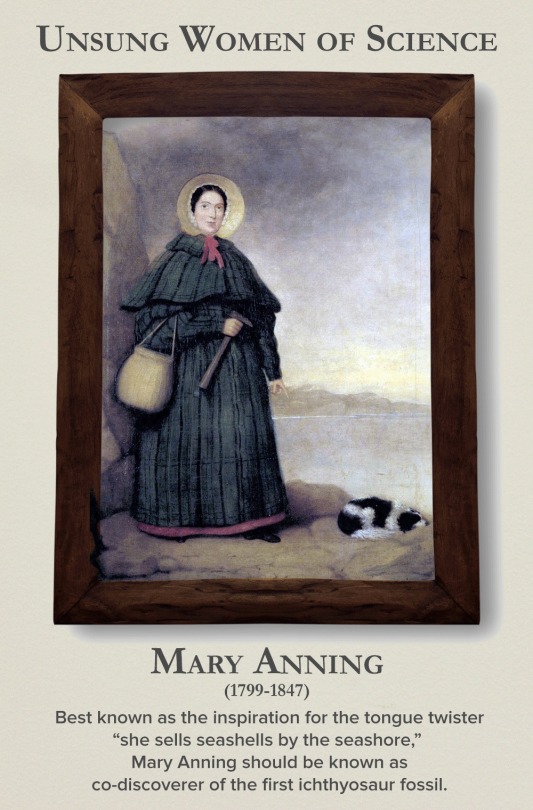

Today is the International Day of Women and Girls in Science, so let’s write women back into science history. Check out the gallery here.
Are colour-changing octopuses really colourblind?
Cephalopods, including octopuses and squid, have some of the most incredible colour-changing abilities in nature.
They can almost instantly blend in with their surroundings to evade predators or lay in wait, and put on colourful displays to attract mates or dazzle potential prey.
This is impressive enough on its own, but becomes even more amazing when you discover these creatures are in fact colourblind – they only have one type of light receptor in their eyes, meaning they can only see in black and white.
So how do they know what colours to change to at all?
This has puzzled biologists for decades but a father/son team of scientists from the University of California, Berkeley, and Harvard University think the unusual shape of their pupils holds the key, and they can see colour after all.
Cephalopods have wide U-shaped or dumbbell-shaped pupils, which allow light into the lens from many directions.

When light enters the pupils in human eyes it gets focused on one spot, cutting down on blur from the light being split into its constituent colours.
The scientists believe cephalopod eyes work the opposite way – the wide pupils split the light up and then individual colours can be focused on the retina by changing the depth of the eyeball and moving the pupil around.
The price for this is blurry vision, but it does mean they could make out colours in a unique way to any other animals.
Processing colour this way is more computationally intensive than other types of colour vision and likely requires a lot of brainpower, which might explain in part why cephalopods are the most intelligent invertebrates on Earth.

Read the paper
Images: Roy Caldwell, Klaus Stiefel, Alexander Stubbs

How are elements created in space, stars, and in laboratories? The latest edition of #PeriodicGraphics in C&EN takes a look! http://bit.ly/2UXWoPD http://bit.ly/2YbuBNE

We Just Moved One Step Closer to Understanding (and Defeating) Alzheimer’s
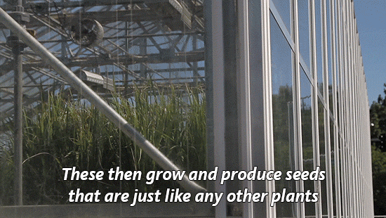
Why we need GMOs to survive climate change
Genetically modified organisms get a bad rap for many reasons, but we’ve actually been genetically altering what we eat since the dawn of human history.
“For 10,000 years, we have altered the genetic makeup of our crops,”explains UC Davis plant pathology professor Pamela Ronald.
“Today virtually everything we eat is produced from seeds that we have genetically altered in one way or another.” (You can read more about Ronald’s thoughts on genetically engineered food here.)
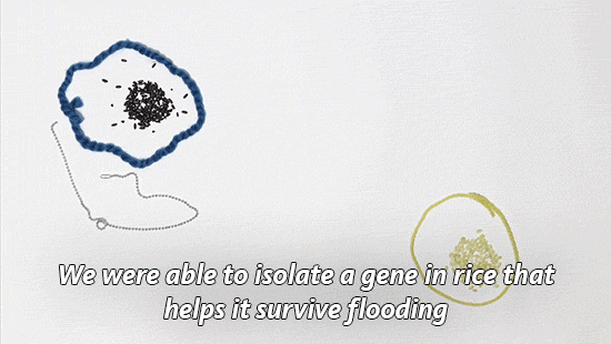
Right now her focus is on rice. It’s one of our basic crops and without it, we would struggle to feed much of the world.
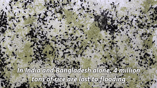
With climate change, we’re seeing an increase in flooding in places like India and Bangladesh, which makes it harder to grow this important food staple.
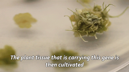
So Ronald and her lab have developed a flood-tolerant strain of rice. It’s known as Sub1a or “scuba rice” and millions of farmers in South Asia are now growing it in their fields.

Today is National Food Day, a day dedicated to hunger awareness. But as we focus on food insecurity, we need to talk more about how global warming will make the problem worse.
As our climate continues to heat up, it has huge impacts on what foods we are able to grow. Will our crops be able to survive droughts and floods? The University of California leads six labs that are working to develop other climate-resilient crops including chickpea, cowpea and millet.
Find out what other scientists are doing to improve our food.

(Image caption: Antidepressants move G proteins out of lipid rafts in the cell membrane. Credit: Molly Huttner)
Why do antidepressants take so long to work?
An episode of major depression can be crippling, impairing the ability to sleep, work, or eat. In severe cases, the mood disorder can lead to suicide. But the drugs available to treat depression, which can affect one in six Americans in their lifetime, can take weeks or even months to start working.
Researchers at the University of Illinois at Chicago have discovered one reason the drugs take so long to work, and their finding could help scientists develop faster-acting drugs in the future. The research was published in the Journal of Biological Chemistry.
Neuroscientist Mark Rasenick of the UIC College of Medicine and colleagues identified a previously unknown mechanism of action for selective serotonin reuptake inhibitors, or SSRIs, the most commonly prescribed type of antidepressant. Long thought to work by preventing the reabsorption of serotonin back into nerve cells, SSRIs also accumulate in patches of the cell membrane called lipid rafts, Rasenick observed, and the buildup was associated with diminished levels of an important signal molecule in the rafts.
“It’s been a puzzle for quite a long time why SSRI antidepressants can take up to two months to start reducing symptoms, especially because we know that they bind to their targets within minutes,” said Rasenick, distinguished professor of physiology and biophysics and psychiatry at UIC. “We thought that maybe these drugs have an alternate binding site that is important in the action of the drugs to reduce depressive symptoms.”
Serotonin is thought to be in short supply in people with depression. SSRIs bind to serotonin transporters – structures embedded within nerve-cell membranes that allow serotonin to pass in and out of the nerve cells as they communicate with one another. SSRIs block the transporter from ferrying serotonin that has been released into the space between neurons – the synapse – back into the neurons, keeping more of the neurotransmitter available in the synapse, amplifying its effects and reducing symptoms of depression.
Rasenick long suspected that the delayed drug response involved certain signaling molecules in nerve-cell membranes called G proteins.
Previous research by him and colleagues showed that in people with depression, G proteins tended to congregate in lipid rafts, areas of the membrane rich in cholesterol. Stranded on the rafts, the G proteins lacked access to a molecule called cyclic AMP, which they need in order to function. The dampened signaling could be why people with depression are “numb” to their environment, Rasenick reasoned.
In the lab, Rasenick bathed rat glial cells, a type of brain cell, with different SSRIs and located the G proteins within the cell membrane. He found that they accumulated in the lipid rafts over time — and as they did so, G proteins in the rafts decreased.
“The process showed a time-lag consistent with other cellular actions of antidepressants,” Rasenick said. “It’s likely that this effect on the movement of G proteins out of the lipid rafts towards regions of the cell membrane where they are better able to function is the reason these antidepressants take so long to work.”
The finding, he said, suggests how these drugs could be improved.
“Determining the exact binding site could contribute to the design of novel antidepressants that speed the migration of G proteins out of the lipid rafts, so that the antidepressant effects might start to be felt sooner.”
Rasenick already knows a little about the lipid raft binding site. When he doused rat neurons with an SSRI called escitalopram and a molecule that was its mirror image, only the right-handed form bound to the lipid raft.
“This very minor change in the molecule prevents it from binding, so that helps narrow down some of the characteristics of the binding site,” Rasenick said.
-
 theoreticallypunkrock reblogged this · 7 years ago
 -
 toonvailo reblogged this · 8 years ago
 -
 atomsandcoffee-blog liked this · 8 years ago
 -
 alixe-studies reblogged this · 8 years ago
 -
 empress-93 reblogged this · 8 years ago
 -
 hitlerfish reblogged this · 8 years ago
 -
hitlerfish liked this · 8 years ago
-
 jas-i-ca-blog reblogged this · 8 years ago
 -
 koki-koki reblogged this · 8 years ago
 -
 moonlightrisesblog liked this · 8 years ago
 -
 yrty123-blog liked this · 8 years ago
 -
 kotonechan32 liked this · 8 years ago
 -
 stormsandwaves liked this · 8 years ago
 -
want-for-rage reblogged this · 8 years ago
-
 vakarianshadow liked this · 8 years ago
 -
 ecolivingbiologist-blog reblogged this · 8 years ago
 -
alixe-studies liked this · 8 years ago
-
 hunted-valkyrie liked this · 8 years ago
 -
 id-be-thor liked this · 8 years ago
 -
 edwordsmyth liked this · 8 years ago
 -
 chibisatou liked this · 8 years ago
 -
 lil-fandomqueen-blog liked this · 8 years ago
 -
 panda-poes reblogged this · 8 years ago
 -
 fatalistic-femme liked this · 8 years ago
 -
 macgyvermedical liked this · 8 years ago
 -
 aninhaelric liked this · 8 years ago
 -
 triapus liked this · 8 years ago
 -
 guavaprincess liked this · 8 years ago
 -
 gigglebitch1996 reblogged this · 8 years ago
 -
 doctor-captain-blog1 reblogged this · 8 years ago
 -
 silentgong reblogged this · 8 years ago
 -
 nerdyneko265 liked this · 8 years ago
 -
 princeofnobody reblogged this · 8 years ago
 -
 emperoronion liked this · 8 years ago
 -
 eyeimagery liked this · 8 years ago
 -
 corprus reblogged this · 8 years ago
 -
 aurartemisa liked this · 8 years ago
A pharmacist and a little science sideblog. "Knowledge belongs to humanity, and is the torch which illuminates the world." - Louis Pasteur
215 posts