Neuroscientists’ Study Sheds Light On How Words Are Represented In The Brain
Neuroscientists’ Study Sheds Light on How Words Are Represented in the Brain
Reading is a relatively modern and uniquely human skill. For this reason, visual word recognition has been a puzzle for neuroscientists because the neural systems responsible for reading could not have evolved for this purpose. “The existence of brain regions dedicated to reading has been fiercely debated for almost 200 years,” said Avniel Ghuman, an assistant professor in the University of Pittsburgh Department of Neurological Surgery. “Wernicke, Dejerine, and Charcot, among the most important and influential neurologists and neuroscientists of the 19th century, debated whether or not there was a visual center for words in the brain.”

In recent years, much of this debate has centered on the left mid-fusiform gyrus, which some call the visual word form area. A recent study by Pitt neuroscience researchers addresses this debate and sheds light on our understanding of the neurobiology of reading.
In a study published July 19 in the Proceedings of the National Academy of Sciences, Ghuman, Elizabeth Hirshorn of Pitt’s Learning Research and Development Center (LRDC), and colleagues from the Department of Psychology and Center for the Neural Basis of Cognition used direct neural recordings and brain stimulation to study the role of the visual word form area in reading in four epileptic patients. The patients chose surgical treatment for their drug-resistant epilepsy and volunteered to participate in the research study. As part of the surgical treatment, neurosurgeons implanted electrodes in the patients’ visual word form area, providing an unprecedented opportunity to understand how the brain recognizes printed words.
First, painless electrical brain stimulation was used through the electrodes to disrupt the normal functioning of the visual word form area, which adversely affected the patients’ ability to read words. One patient dramatically misperceived letters, and another felt that there were words and parts of words present that were not in what she was reading. Stimulation to this region did not disrupt their ability to name objects or faces. A brief video of the stimulation can be seen here.
In addition to stimulating through these electrodes, the activity from the area was recorded while the patients read words. Using techniques from machine learning to analyze the brain activity that evolved over a few hundred milliseconds from this region, the researchers could tell what word a patient was reading at a particular moment. This suggests that neural activity in the area codes knowledge about learned visual words that can be used to discriminate even words that are only one letter different from one another (for example, “hint” and “lint”).
“This study shows that the visual word form area is exquisitely tuned to the fine details of written words and that this area plays a critical role in refining the brain’s representation of what we are reading. The disrupted word and letter perception seen with stimulation provides direct evidence that the visual word form area plays a dedicated role in skilled reading,” said Hirshorn. “These results also have important implications for understanding and treating reading disorders. The activity in the visual word form area, along with its interactions with other brain areas involved in language processing, could be a marker for proficient reading. Having a better understanding of this neural system could be critical for diagnosing reading disorders and developing targeted therapies.”
“It is exciting that with modern brain-recording techniques and advanced analysis methods, we are finally able to start answering questions about the brain and the mind that people have asked for centuries and contribute to our understanding of reading disorders,” said Ghuman.
More Posts from Contradictiontonature and Others
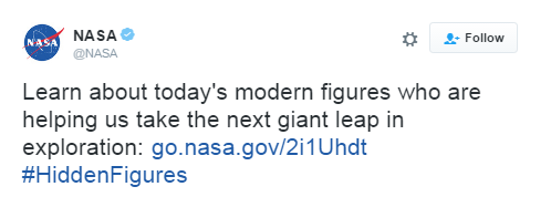
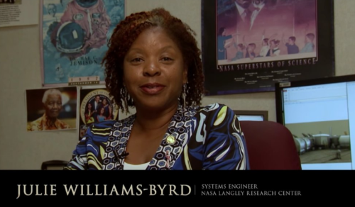
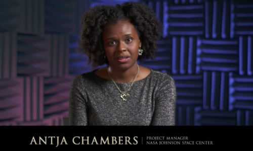

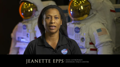
The film “Hidden Figures,” based on the book by Margot Lee Shetterly, focuses on the stories of Katherine Johnson (left, after receiving the Medal of Freedom in 2015), Mary Jackson and Dorothy Vaughan, African-American women who were essential to the success of early spaceflight. Today, NASA embraces their legacy and strives to include everyone who wants to participate in its ongoing exploration. “Progress is driven by questioning our assumptions and cultural assumptions,” NASA Administrator Charles Bolden says in a new video. “Embracing diversity and inclusion is how we as a nation will take the next giant leap in exploration.“
- Source
Let’s learn about today’s black heroes we all can look up to!
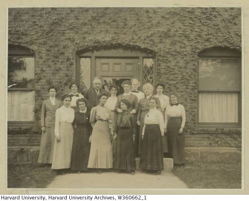
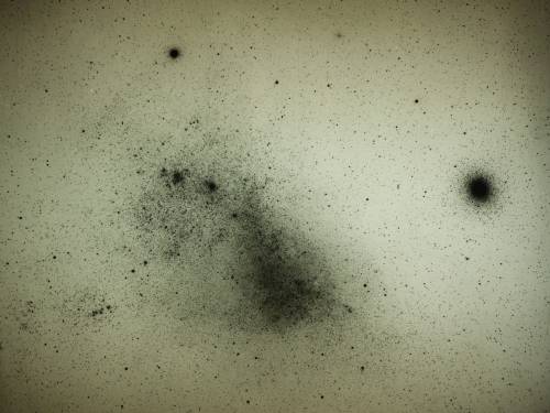
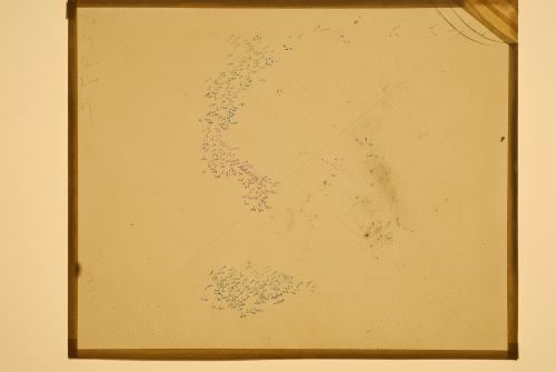
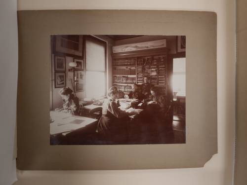
This team of early female astronomers created the star classification system we use today.
In the late 19th century, astronomy was a growing field. At the time, Edward Pickering, the director of the Harvard College Observatory, was working to create a classification system for stars by capturing the light from these distant celestial objects onto photographic glass plates. A team of female assistants and astronomers meticulously maintained and analyzed these delicate negatives. In her new book, The Glass Universe: How the Ladies of the Harvard Observatory Took the Measure of the Stars, Dava Sobel shares the stories of these female “human computers” and how their work helped to advance the field of astronomy and the role of women in science.
This team of astronomers included Williamina Fleming, who was once Pickering’s maid but eventually became a supervisor to the group and went on to identify hundreds of variable stars. And Henrietta Swan Leavitt’s observations about the luminosity of stars would shape later ideas about the expanding universe.
Listen to the interview here.
[Photos courtesy of The Glass Universe]

Popcorn’s explosive pop looks pretty cool in high-speed video, but just watching it with a regular camera doesn’t show everything that’s going on. If we take a look at it through schlieren optics, the kernel’s pop looks even more extraordinary:

The schlieren technique reveals density differences in the gases around the corn–effectively allowing us to see what is invisible to the naked eye. The popcorn kernel acts like a pressure vessel until the expansion of steam inside causes its shell to rupture. The first hints of escaping steam send droplets of oil shooting upward. The kernel may hop as steam pours out the rupture point, causing the turbulent billowing seen in the animation above. As the heat causes legs of starch to expand out of the kernel, they can push off the ground and propel the popcorn higher. As for the eponymous popping sound, that is the result of escaping water vapor, not the actual rupture or rebound of the kernel! See more of the invisible world surrounding a popping kernel in the video below. (Image credits: Warped Perception, source; Bell Labs Ireland, source; WP video via Gizmodo; BLI video submitted by Kevin)


TOP TEN MOST DEADLY INFECTIOUS DISEASES
This list is based off of the assumption that the infected individual does not receive medical treatment.
1. Prions (mad cow disease, Creutzfeld-Jakob disease, kuru, fatal familial insomnia): 100%
2. Rabies: ~100%
3. African trypanosomiasis (’African sleeping sickness’): ~100%
4. Primary amoebic encephalitis caused by Naegleri fowlerii (’the brain-eating amoeba’): ~100%
5. Yersinia pestis, specifically the pneumonic or septicemic subtype (’the black plague’): ~100%
6. Visceral leishmaniasis: ~100%
7. Smallpox, specifically the malignant (flat) or hemorragic subtype: 95%
8. Ebola virus, specifically the Zaire strain: 83-90%
9. HIV: 80-90%
10. Anthrax, specifically the pulmonary subtype: >85%

If you dropped a water balloon on a bed of nails, you’d expect it to burst spectacularly. And you’d be right – some of the time. Under the right conditions, though, you’d see what a high-speed camera caught in the animation above: a pancake-shaped bounce with nary a leak. Physically, this is a scaled-up version of what happens to a water droplet when it hits a superhydrophobic surface.
Water repellent superhydrophobic surfaces are covered in microscale roughness, much like a bed of tiny nails. When the balloon (or droplet) hits, it deforms into the gaps between posts. In the case of the water balloon, its rubbery exterior pulls back against that deformation. (For the droplet, the same effect is provided by surface tension.) That tension pulls the deformed parts of the balloon back up, causing the whole balloon to rebound off the nails in a pancake-like shape. For more, check out this video on the student balloon project or the original water droplet research. (Image credits: T. Hecksher et al., Y. Liu et al.; via The New York Times; submitted by Justin B.)

Watching a snowflake grow seems almost magical–the six-sided shape, the symmetry, the way every arm of it grows simultaneously. But it’s science that guides the snowflake, not magic. Snowflakes are ice crystals; their six-sided shape comes from how water molecules fit together. The elaborate structures and branches in a snowflake are the result of the exact temperature and humidity conditions when that part of the snowflake formed. The crystals look symmetric and seem to grow identical arms simultaneously because the temperature and humidity conditions are the same around the tiny forming crystals. And the old adage that no two snowflakes are alike doesn’t hold either. If you can control the conditions well enough, you can grow identical-twin snowflakes! (Video credit: K. Libbrecht)

This massive virus with its own immune system could hold the future of medicine
French researchers think they’ve found a giant virus big enough to house its own virus-killing devices using a system like CRISPR, and it could be a completely new form of life.
Called a mimivirus, it was first found growing in amoebae in a water tower. At four times the size of a typical virus, you can even see it under a light microscope
When the mimivirus encounters another virus, it stores some of the invader’s genetic material. That way, when it encounters the same kind of virus again, the MIMIVIRE system goes into gene-editing berserker mode, finding the key genes of the virus and cutting them to inert oblivion. This could have major applications.
Follow @the-future-now

Viruses support photosynthesis in bacteria: An evolutionary advantage?
Viruses propagate by infecting a host cell and reproducing inside. This not only affects humans and animals, but bacteria as well. This type of virus is called bacteriophage. They carry so called auxiliary metabolic genes in their genome, which are responsible for producing certain proteins that give the virus an advantage. Researchers at the University of Kaiserslautern and the Ruhr University Bochum have analysed the structure of such a protein more closely. It appears to stimulate the photosynthesis of host bacteria. The study has now been published in the journal The Journal of Biological Chemistry.
Raphael Gasper, Julia Schwach, Jana Hartmann, Andrea Holtkamp, Jessica Wiethaus, Natascha Riedel, Eckhard Hofmann, Nicole Frankenberg-Dinkel. Auxiliary metabolic genes- Distinct Features of Cyanophage-encoded T-type Phycobiliprotein Lyase θCpeT. Journal of Biological Chemistry, 2017; jbc.M116.769703 DOI: 10.1074/jbc.M116.769703
The association between the virus protein and bacterial pigment is incredibly stable. Furthermore, the complex is highly fluorescent. Credit: AG Frankenberg-Dinkel


A special organic dye, Nile Red in different solvents.
From left to right I dissolved equal amounts of Nile Red (a dye) in different solvents. The solvents were: methanol, diisopropyl ether, hexane, n-propanol, tetrahydrofuran, toluene, ethanol, acetone.
Depending on the solvents polarity, the dye dissolved to give different colored solutions (upper image), this is called solvatochromism. It is the ability of a chemical substance to change color due to a change in solvent polarity.
Under UV light, these solutions emitted different colors (bottom pics), this is called solvatofluorescence. The emission and excitation wavelength both shift depending on solvent polarity, so it fluoresces with different color depending on the solvent what it’s dissolved in.
Nile Red is a quite expensive dye, which costs a bit over 1000 USD/gram, therefore I had to make it. The purification of the raw material was posted HERE.
To help the blog, donate to Labphoto through Patreon: https://www.patreon.com/labphoto


Dancing Flames - Aluminium’s reactivity
aluminium’s protective oxide layer can make it difficult to see its true reactivity in the context of metals reacting with aqueous solutions. (Do not try to recreate experiment without the presence of a trained professional)
Procedure:
Dissolve copper(II) chloride in the hydrochloric acid and set the solution aside. Take a piece of aluminium foil approximately the width of the base of the conical flask and approximately 20 cm long. Roll it loosely just enough to be able to fit through the neck of the flask – use a splint or spatula to gently push it home so it lies on its side on the base of the flask. Have a source of ignition nearby with some splints. Pour the solution inside the flask and swirl the flask gently to get the reaction going.
After a few seconds, the mixture will begin to react vigorously and produce hydrogen gas. Hold the lit splint by the opening of the flask and the gas will ignite. If you have timed it right, the flame will sink back into the flask and dance inside above the reaction with an eerie green color from the copper.
Reaction:
2Al(s) + 3CuCl2(aq) → 2AlCl3(aq) + 3Cu(s)
2Al(s) + 6HCl(aq) → 2AlCl3(aq) + 3H2(g)
The primary objective here is to show how reactive aluminium is. The aluminium oxide layer protects the metal beneath from further reaction with air, water or acid. But chloride ions can ligate aluminium ions at the metal–oxide interface and break down the protective layer, allowing the reaction to proceed. -rsc
Giffed by: rudescience From: This video
-
 iforgetoneartheat liked this · 7 years ago
 -
 czkavka reblogged this · 7 years ago
 -
 re-wiredbrain-blog liked this · 8 years ago
 -
 gold-34-heart liked this · 8 years ago
 -
 lowdecembersun reblogged this · 8 years ago
 -
 nonbinary-in-stem-blog reblogged this · 8 years ago
 -
 praziquantelheartburn reblogged this · 8 years ago
 -
 onlyobeythedaleks reblogged this · 8 years ago
 -
 1grammiefan liked this · 8 years ago
 -
 shinigamia liked this · 8 years ago
 -
 jeenay liked this · 8 years ago
 -
 mangoachaar reblogged this · 8 years ago
 -
 kayura-fii liked this · 8 years ago
 -
 floutitude liked this · 8 years ago
 -
 beauvale reblogged this · 8 years ago
 -
beauvale liked this · 8 years ago
-
 whatisthisreally liked this · 8 years ago
 -
 nessacul liked this · 8 years ago
 -
 mysterysciencegirlfriend3000 reblogged this · 8 years ago
 -
 queenofeire liked this · 8 years ago
 -
 mullercells reblogged this · 8 years ago
 -
mullercells liked this · 8 years ago
-
 coolhamdi reblogged this · 8 years ago
 -
 fancygremlin reblogged this · 8 years ago
 -
 narcisphd reblogged this · 8 years ago
 -
narcisphd liked this · 8 years ago
-
 infodump-playhouse reblogged this · 8 years ago
 -
 infiniteglitterfall liked this · 8 years ago
 -
 normanborblaug reblogged this · 8 years ago
 -
 turtletotem liked this · 8 years ago
 -
 bananaseven reblogged this · 8 years ago
 -
bananaseven liked this · 8 years ago
-
 crystals-and-catalysis-blog liked this · 8 years ago
 -
 an-aerosol-mist-of-blood liked this · 8 years ago
 -
 livingtospitetheangst liked this · 8 years ago
 -
 euphorbic reblogged this · 8 years ago
 -
 brittaniahart liked this · 8 years ago
 -
 contradictiontonature reblogged this · 8 years ago
A pharmacist and a little science sideblog. "Knowledge belongs to humanity, and is the torch which illuminates the world." - Louis Pasteur
215 posts