The Film “Hidden Figures,” Based On The Book By Margot Lee Shetterly, Focuses On The Stories Of Katherine
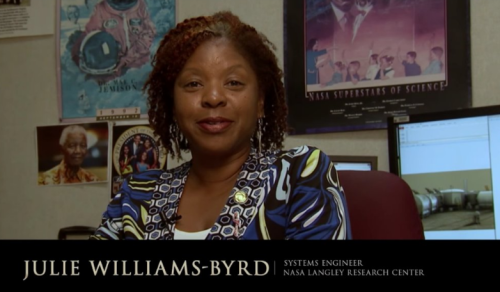
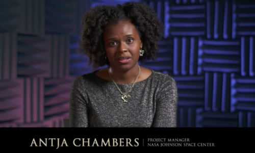

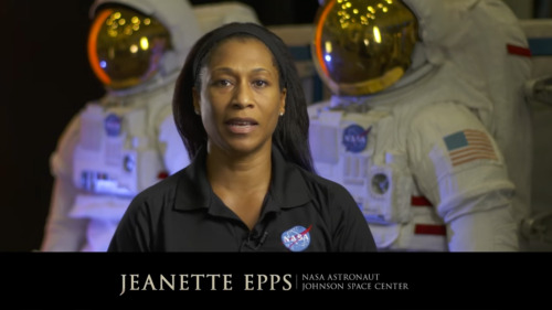
The film “Hidden Figures,” based on the book by Margot Lee Shetterly, focuses on the stories of Katherine Johnson (left, after receiving the Medal of Freedom in 2015), Mary Jackson and Dorothy Vaughan, African-American women who were essential to the success of early spaceflight. Today, NASA embraces their legacy and strives to include everyone who wants to participate in its ongoing exploration. “Progress is driven by questioning our assumptions and cultural assumptions,” NASA Administrator Charles Bolden says in a new video. “Embracing diversity and inclusion is how we as a nation will take the next giant leap in exploration.“
- Source
Let’s learn about today’s black heroes we all can look up to!
More Posts from Contradictiontonature and Others


My new favorite: Solvatofluorescence of Nile Red
Solvatochromism is the ability of a chemical substance to change color due to a change in solvent polarity, so it has different color in different solvents.
Also in some cases, the emission and excitation wavelength both shift depending on solvent polarity, so it fluoresces with different color depending on the solvent what it’s dissolved in. This effect is solvatofluorescence.
On the video the highly solvatochromic organic dye, Nile Red was added to different organic solvents and was diluted with another, different polarity organic solvent. As the polarity of the solution changed, the emitted color from the fluorescent dye also varied as seen on the gifs above and as seen on the video:
To help the blog, donate to Labphoto through Patreon: https://www.patreon.com/labphoto

IN THE MIX
Matti Koivisto, an undergraduate student working in the laboratory of Kari Haajanen at Turku University of Applied Sciences, designs solutions similar to this one to detect the bacteria Escherichia coli. He uses a dye called rhodamine 6G, which has a strong orange color when dissolved in ethanol. In solution, the dye separates into negatively and positively charged ions, the latter of which are largely responsible for the dye’s color. Negatively charged molecules on the outer membranes of E. coli attract the dye’s positive ions. This interaction causes the dye to change color to a pinkish hue, and the level of color change allows Koivisto to gauge how many of the bacteria are present.
Submitted by Matti Koivisto
Do science. Take photos. Make money: Enter our monthly photo contest here for your chance to win!
Related C&EN content:
How dyes in mouse feces help track insects
Using an ink-jet printer to simplify analytical chemistry


COLORS OF CHEMISTRY
The bright colors of chemistry fascinate people of all ages. Hriday Bhattacharjee, a Ph.D. student in the lab of Jens Mueller at the University of Saskatchewan, assembled this showcase from compounds he prepared as well as from some synthesized by the undergraduate students he teaches. Organometallic and inorganic chemistry—the study of molecules like these that involve metal atoms—is especially colorful.
The table below the picture indicates the chemicals seen in the photo.
Submitted by Hriday Bhattacharjee
Do science. Take pictures. Win money: Enter our photo contest.

This Week in Chemistry: Preventing marble statue weathering, further progress towards hydrogen fusion, and more! Links: http://goo.gl/WeJRV5
Neil deGrasse Tyson talking about creationism, science celebrities and kids on National Geographic. Watch the full video here.


Feeling a little small? Well in the context of the cosmos, we are small. We may just be little guys living on a speck of dust, afloat in a staggering immensity…
…but we dont think small.

What Happens in the Brain During Unconsciousness?
Researchers are shining a light on the darkness of the unconscious brain. Three new studies add to the body of knowledge.
When patients undergo major surgery, they’re often put under anesthesia to allow the brain to be in an unconscious state.
But what’s happening in the brain during that time?
Three Michigan Medicine researchers are authors on three new articles from the Center for Consciousness Science exploring this question — specifically how brain networks fragment in association with a variety of unconsciousness states.
“These studies come from a long-standing hypothesis my colleagues and I have had regarding the essential characteristic of why we are conscious and how we become unconscious, based on patterns of information transfer in the brain,” says George A. Mashour, M.D., Ph.D., professor of anesthesiology, director of the Center for Consciousness Science and associate dean for clinical and translational research at the University of Michigan Medical School.
In the studies, the team not only explores how the brain networks fragment, but also how better to measure what is happening.
“We’ve been working for a decade to understand in a more refined way how the spatial and temporal aspects of brain function break down during unconsciousness, how we can measure that breakdown and the implications for information processing,” says UnCheol Lee, Ph.D., physicist, assistant professor of anesthesiology and associate director of the Center for Consciousness Science.
Examining different aspects of unconsciousness
The basis for the three studies, as well as other work from the Center for Consciousness Science, comes from a theory Mashour produced during his residency.
“I published a theoretical article when I was a resident in anesthesiology suggesting that anesthesia doesn’t work by turning the brain off, per se, but rather by isolating processes in certain areas of the brain,” Mashour says. “Instead of seeing a highly connected brain network, anesthesia results in an array of islands with isolated cognition and processing. We have taken this thought, as well as the work of others, and built upon it with our research.”
In the study in the Journal of Neuroscience, the team analyzed different areas of the brain during sedation, surgical anesthesia and a vegetative state.
“It’s often suggested that different areas of the brain that typically talk to one another get out of sync during unconsciousness,” says Anthony Hudetz, Ph.D., professor of anesthesiology, scientific director of the Center for Consciousness Science and senior author on the study. “We showed in the early stages of sedation, the information processing timeline gets much longer and local areas of the brain become more tightly connected within themselves. That tightening might lead to the inability to connect with distant areas.”
In the Frontiers in Human Neuroscience study, the team delved into how the brain integrates information and how it can be measured in the real world.
“We took a very complex computational task of measuring information integration in the brain and broke it down into a more manageable task,” says Lee, senior author on the study. “We demonstrated that as the brain gets more modular and has more local conversations, the measure of information integration starts to decrease. Essentially, we looked at how the brain network fragmentation was taking place and how to measure that fragmentation, which gives us the sense of why we lose consciousness.”
Finally, the latest article, in Trends in Neurosciences, aimed to take the team’s previous studies and other work on the subject of unconsciousness and put together a fuller picture.
“We examined unconsciousness across three different conditions: physiological, pharmacological and pathological,” says Mashour, lead author on the study. “We found that during unconsciousness, disrupted connectivity in the brain and greater modularity are creating an environment that is inhospitable to the kind of efficient information transfer that is required for consciousness.”
How these studies can help patients
The team members at the Center for Consciousness Science note that all of this work may help patients in the future.
“We’re looking for a better way to quantify the depth of anesthesia in the operating room and to assess consciousness in someone who has had a stroke or brain damage,” Hudetz says. “For example, we may assume that a patient is fully unconscious based on behavior, but in some cases consciousness can persist despite unresponsiveness.”
The team hopes this and future research could lead to therapeutic strategies for patients.
“We want to understand the communication breakdown that occurs in the brain during unconsciousness so we can precisely target or monitor these circuits to achieve safer anesthesia and restore these circuits to improve outcomes of coma,” Mashour says.


Heart Regeneration
If a person suffers a heart attack and survives, chances are their heart muscle will never be quite the same. Indeed, the associated scarring often results in permanent damage that can lead to heart failure and eventual death. Scientists are therefore searching for possible ways to promote regeneration of damaged hearts, and it’s possible that newborn mice may hold the answer. For a few weeks after birth, these animals can almost entirely regenerate their heart tissue after an injury. And new research suggests a key process that may be critical for this regenerative ability: regrowth of nerves. Blocking nerve growth specifically in experimental animals completely prevented the regrowth of damaged heart tissue. In control animals, the nerves regrew into their normal branching patterns—like those pictured. Thus if researchers are to have any hope of regenerating adult hearts after injury, their best bet might be to boost accompanying nerve growth.
Written by Ruth Williams
Image from work by Ian A. White and colleagues
Interdisciplinary Stem Cell Institute, University of Miami Miller School of Medicine, USA
Image copyright held by the American Heart Association
Published in Circulation Research, December 2015
You can also follow BPoD on Twitter and Facebook
Dive deep into Episode 05 of #ShelfLife to discover the various technologies that have helped humans map the sky around us for eons.

From sundials to mega-powered modern telescopes, tools for stargazing allow us to understand the universe—and our place within it. Season 2 begins on November 1.

-
 itzmellowmeow liked this · 4 months ago
 -
 fox-of-glass liked this · 1 year ago
 -
 getbacktofandomlife reblogged this · 2 years ago
 -
 jeanp-blog liked this · 4 years ago
 -
 belovedbluebells liked this · 4 years ago
 -
 graveltoglucose liked this · 4 years ago
 -
 baklava-yandere reblogged this · 4 years ago
 -
baklava-yandere liked this · 4 years ago
-
 kiss-me-sober liked this · 5 years ago
 -
 jjneeps liked this · 5 years ago
 -
 to-be-fit-or-not-to-be-fit reblogged this · 5 years ago
 -
 20sidedfitness liked this · 5 years ago
 -
 scrumptiouslyunknown reblogged this · 5 years ago
 -
 talk-shit-get-fit reblogged this · 5 years ago
 -
 icallcurrency reblogged this · 5 years ago
 -
 starryknight63 liked this · 5 years ago
A pharmacist and a little science sideblog. "Knowledge belongs to humanity, and is the torch which illuminates the world." - Louis Pasteur
215 posts