It’s Nearly The Season Of The Witch: Time For Some Cauldron Chemistry

It’s nearly the season of the witch: time for some cauldron chemistry
Midwives and nurses sometimes came under suspicion because of their specialised knowledge and success - or failure - in treating those who were sick. These healing roles were traditionally taken on by women who, until around the turn of the nineteenth century, were excluded from formal medical training. However, many still practiced medicine in their homes and villages, and what they had learned came from shared knowledge and trial and error, rather than accepted official sources. A medical education might not have been a great help in any case. In the days before germ theory the causes of sickness and the reasons for recovery were not obvious. Any recoveries could be seen as miraculous … or the result of witchcraft.
Treating sickness and disease pre-germ theory was largely guesswork. All sorts of noxious compounds were administered to ailing individuals, and if they produced any effect on the body, be it vomiting, diarrhoea or sweating, it was seen as a good thing – and that was the practice of the so-called professionals. It is not hard to see how images of unofficial healers and herbalists (both men and women) stooping over boiling pots of herbs, roots and who-knows-what, could become a template for the image of a witch, especially when many of the concoctions they produced had such unpleasant effects on their patients.
Having said that, herbalists and traditional healers should not be dismissed as completely ignorant of the medical benefits of some of the plants and poultices they used. Some of the ingredients associated with traditional healing and witches’ potions have been found to be hugely beneficial to medicine once they have been isolated, tested and modified. Science has enabled us to identify the key components of some plants and test them to determine how and when they should be administered safely and effectively. Chemists have modified the structures of some of the compounds to reduce side-effects, make drugs more potent or lower their toxicity.
More Posts from Contradictiontonature and Others

The 2016 Nobel Prize in Chemistry is awarded to Jean-Pierre Sauvage, Sir Fraser Stoddart, and Bernard Feringa for the design and production of molecular machines with controllable movements: bit.ly/NobelSci2016
Most cones don’t really see color
We see color because of specialized light-sensing cells in our eyes called cones. One type, L-cones, sees the reds of strawberries and fire trucks; M-cones detect green leaves, and S-cones let us know the sky is blue. But vision scientists have now discovered that not all cones sense color (see video). The finding was made possible because, for the first time, scientists were able to look at individual photo-sensing cells.


Maryam Mirzakhani was an Iranian mathematician and a professor of mathematics at Stanford University. She was the first-ever female winner of the prestigious Fields Medal prize and the first Iranian to be honoured with the award.
Mirzakhani was born in Tehran, Iran. She attended Farzanegan School, which was part of the National Organization for Development of Exceptional Talents. In both 1994 and 1995 she won the International Mathematical Olympiads for high-school students. In the 1995 International Mathematical Olympiad, she became the first Iranian student to achieve a perfect score and to win two gold medals.
Mirzakhani continued her education at Sharif University of Technology in Tehran, where she earned a BSc in Mathematics. After this, she undertook a a Ph.D. from Harvard University. She worked under the supervision of the Fields Medalist Curtis T. McMullen, and her dissertation focused on Simple Geodesics on Hyperbolic Surfaces and Volume of the Moduli Space of Curves. She had a unique way of working, and “would spend hours on the floor with supersized canvases of paper, sketching out ideas, drawing diagrams and formulae, often leading Anahita [her daughter] to say, “Oh, Mommy is painting again!” Mirzankhani said that “I don’t have any particular recipe [for developing new proofs] … It is like being lost in a jungle and trying to use all the knowledge that you can gather to come up with some new tricks, and with some luck you might find a way out.”
From 2004 to 2008 she was a Clay Mathematics Institute Research Fellow and an assistant professor at Princeton University. She then became a professor at Stanford University where she specialized in theoretical mathematics including moduli spaces, Teichmüller theory, hyperbolic geometry, Ergodic theory and symplectic geometry.”
In 2014, Mirzakhani was awarded the Fields Medal prize for her work on complex geometry and dynamic systems, becoming the first-ever female winner and the first Iranian to be honoured with the award. During her lifetime, she won a number of awards including the 2009 Blumenthal Award for the Advancement of Research in Pure Mathematics and the 2013 Satter Prize of the American Mathematical Society. She worked up until her death in 2017, and was still producing amazing mathematics during her battle with cancer over the last few years.
Sources here, here, here, here and here
Antibiotic Resistance Will Soon Hit the Tipping Point, Unless We Act
Antibiotic Resistance Will Soon Hit the Tipping Point, Unless We Act
Antibiotic-resistant superbugs are enough of a severe, genuine threat to global populations that the UN has placed the issue on par with the spread of Ebola and HIV. The livestock industry is a major factor contributing to the rapid proliferation of these superbugs, swift action is required.
Liver Circulation - Flashcard

The liver is supplied with blood by the hepatic artery and the hepatic portal vein
branches of the hepatic artery and the hepatic portal vein distribute blood to the periphery of the liver lobules.
Blood passes along sinusoids, which are lined by hepatocytes, which perform numerous metabolic and synthetic functions.
The processed blood passes into branches of the hepatic vein in the centre of each lobule, and eventually drains into the hepatic vein.
The biliary system is independent of the vascular system and bile moves in the opposite direction to the blood.
Initially it is collected in bile ductules which are surrounded by collagenous tissue, which forms part of the collagenous trabecular septum.
The bile is collected by increasingly large trabecular ducts, which fuse to form intrahepatic ducts which finally drain into the main hepatic ducts.


Antibiotic resistance has been called one of the biggest public health threats of our time. There is a pressing need for new and novel antibiotics to combat the rise in antibiotic-resistant bacteria worldwide.
Researchers from Florida International University’s Herbert Wertheim College of Medicine are part of an international team that has discovered a new broad-spectrum antibiotic that contains arsenic. The study, published in Nature’s Communication Biology, is a collaboration between Barry P. Rosen, Masafumi Yoshinaga, Venkadesh Sarkarai Nadar and others from the Department of Cellular Biology and Pharmacology, and Satoru Ishikawa and Masato Kuramata from the Institute for Agro-Environmental Sciences, NARO in Japan.
“The antibiotic, arsinothricin or AST, is a natural product made by soil bacteria and is effective against many types of bacteria, which is what broad-spectrum means,” said Rosen, co-senior author of the study published in the Nature journal, Communications Biology. “Arsinothricin is the first and only known natural arsenic-containing antibiotic, and we have great hopes for it.”
Although it contains arsenic, researchers say they tested AST toxicity on human blood cells and reported that “it doesn’t kill human cells in tissue culture.”
Continue Reading.

Autonomic nervous system
Structure and Function of the Sympathetic and Parasympathetic nervous system
The main function of the autonomic nervous system (ANS) is to assist the body in maintaining a relatively constant internal environment. For example, a sudden increase in systemic blood pressure activates the baroreceptors (those are receptors that detect physical pressure) which in turn modify the activity of the ANS so that the blood pressure is restored to its previous level [1].
The ANS is often regarded as a part of the motor system and is responsible for involuntary action and its effector organs are smooth muscle, cardiac muscle and glands. Another system, the somatic (meaning around the body) nervous system, is responsible for voluntary action in which skeletal muscle is the effector.
The ANS can further be divided into 3 parts: sympathetic, parasympathetic and enteric nervous systems [1][2], with the enteric nervous system sometimes being considered a separate entity [2]. Both parasympathetic and sympathetic nervous systems coexist and work in opposition with each other, ultimately maintaining the correct balance; the activity of one being more active depending on the situation. In a normal resting human, the parasympathetic nervous system dominates, while in a tense and stressful situation, the sympathetic nervous system switches to become dominant.

Figure 1. Structure and function of the central nervous system
This article will be focused on sympathetic and parasympathetic activity from the perspective of:
Anatomy
Biochemical
The sympathetic division provides your “fight or flight” whereas the parasympathetic division helps you to “rest and digest”
Anatomy
Higher centers that control autonomic function include the pons, medulla oblongata and hypothalamus [3].
The pons contains the micturition (urination) and respiratory center.
The medulla oblongata contains the respiratory, cardiac, vomiting, vasomotor and vasodilator centres [4].
The hypothalamus contains the highest concentration of autonomic centres [4]. It contains several centres that control autonomic activities, including heat loss, heat production and conservation, feeding and satiety, as well as fluid intake [4].

Figure 2. Locations of the autonomic control centres of the brain
All 3 structures receive input from certain sources by stimulation of nerve fibres resulting from chemical changes in blood composition like blood pH, blood glucose level, blood osmolarity and volume [4]. Notably, the hypothalamus receives input from cerebral cortex and the limbic system, a system that helps control emotional behaviour [3].
Autonomic promoter neurons are neurons that are found in the brain stem, hypothalamus or even cerebral hemispheres that project to preganglionic neurons (discussed below), where they form synapses with these neurons (5). Hence, input from the higher centres can be relayed to the motor neurons (preganglionic and then postganglionic neurons) which subsequently innervate different body tissues. Changes in the input from these centres could result in responses in those tissues.
The primary functional unit of the sympathetic and parasympathetic nervous system consists of a 2 neuron motor pathway (Figure 3), containing a preganglionic and postganglionic neurons, arranged in series.(2) The two synapse in peripheral ganglion. This clearly distinguishes autonomic motor nervous system and somatic nervous system. The somatic nervous system project from the CNS directly to innervated tissue without any intervening ganglia.(6)

Figure 3. Diagram showing the primary functional unit of the ANS
Sympathetic nervous system
Sympathetic preganglionic neurons mainly are concentrated in the lateral horn in the thoracic (T1-12) and upper lumbar (L1 &2) segments of the spinal cord (Figure 4).
The preganglionic axons leave the spinal cord in 3 ways:
Through the paravertebral ganglion
The preganglionic axon may synapse with postganglionic neurons in this ganglion or some axon may travel rostrally or caudally within the sympathetic trunk before forming synapse with a postganglionic neurons in a different paravertebral ganglion.
Through the prevertebral ganglion
Some preganglionic axons pass the paravertebral ganglion (no synapse occur) and form synapse with postganglionic neurons in prevertebral ganglion, also known as collateral ganglion.
Directly to the organs without any synapse
Some preganglionic axons pass through the sympathetic trunk (no synapse) and end directly on cells of the adrenal medulla, which are equivalent to postganglionic cell.
Parasympathetic nervous system
The parasympathetic preganglionic neurons are located in several cranial nerve nuclei in the brain stem and some are found in the S3 and S4 segments of the sacral spinal cord (Figure 4). The parasympathetic postganglionic neurons are located in cranial ganglia, including the ciliary ganglion, the pterygopalatine, submandibular ganglia, and the otic ganglion. Other ganglia are present near or in the walls of visceral organs. Similarly, the preganglionic neurons form synapse with the postganglionic neurons in the ganglia.

Figure 4. Anatomy of the ANS and how its nuerons innervate tissues
After knowing how nerves connect from the CNS to PNS and to different organs, we will now consider some of the neurotransmitters that are being released at different nerve terminals. It is the binding of these neurotransmitters to the receptors on the effectors that leads to biochemical and physiological changes. Some of the neurotransmitters in use are:
For the synapse between pre and postganglionic neurons mentioned above, the neurotransmitter that is released by the preganglionic axon terminal, is acetylcholine. The corresponding receptors are found on the postsynaptic membrane of postganglionic nerves and are nicotinic receptors.
Parasympathetic postganglionic nerve terminals also release acetylcholine.
Sympathetic postganglionic nerve terminals release mostly noradrenaline
The adrenal medulla receives direct stimulation from sympathetic preganglionic innervation, releases mainly adrenaline (80%) and some noradrenaline into the blood stream. In this case, both adrenaline and noradrenaline act as hormones as they are transported via blood circulating system to target organs instead of neuronal pathway.
Strangely, for the sympathetic postganglionic nerves that innervate the sweat glands, the nerves release acetylcholine (normally only by parasympathetic postganglionic nerve) instead.
1. H.P.Rang, J.M.Ritter, R.J.Flower GH. RANG & DALE’S Pharmacology. In: 8th ed. ELSEVIER CHURCHILL LIVINGSTONE; 2016. p. 145.
2. Bruce M. Koeppen BAS. BERNE & LEVY PHYSIOLOGY. In: 6th ed. MOSBY ELSEVIER; 2010. p. 218.
3. Cholinergic transmission [Internet]. 2015. Available from: http://www.dartmouth.edu/~rpsmith/Cholinergic_Transmission.html
4. Bruce M. Koeppen BAS. BERNE & LEVY PHYSIOLOGY. In: 6th ed. MOSBY ELSEVIER; 2016. p. 44.
Thumpety thump thump thumpety thump thump look at Kinesin go
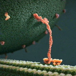
Myosin, kinesin, and dynein are important proteins governing internal transport. Myosin attached to organelles associates with actin microfilaments to enable the continuous flow of cytoplasm called cytoplasmic streaming.
Kinesins and dynein enable the movement of organelles along microtubules. They attach and move along microtubules. Most kinesins transport organelles from the center towards the periphery of the cell, anterograde transport. Dynein, and a few types of kinesins transport towards the cell center, retrograde transport.
(via thelifeofapremed)


Today is the International Day of Women and Girls in Science, so let’s write women back into science history. Check out the gallery here.
-
 natphylo reblogged this · 8 years ago
 -
 littlemariecat reblogged this · 8 years ago
 -
 joyousofthematrix liked this · 8 years ago
 -
 sifilord liked this · 8 years ago
 -
sifilord reblogged this · 8 years ago
-
 scrollingivy reblogged this · 8 years ago
 -
 qravatus liked this · 8 years ago
 -
 spacey-kate reblogged this · 8 years ago
 -
 nmerteuil liked this · 8 years ago
 -
 lucassporl-blog liked this · 8 years ago
 -
luciferapollyon reblogged this · 8 years ago
-
 queenofeire liked this · 8 years ago
 -
 panda-poes reblogged this · 8 years ago
 -
 petit-dino liked this · 8 years ago
 -
 pepiswasp liked this · 8 years ago
 -
 masqueradingfangirl-blog reblogged this · 8 years ago
 -
masqueradingfangirl-blog liked this · 8 years ago
-
spacey-kate liked this · 8 years ago
-
 aninhaelric liked this · 8 years ago
 -
 vinesnweeds liked this · 8 years ago
 -
 bellafuga reblogged this · 8 years ago
 -
 lunacorvum reblogged this · 8 years ago
 -
 treatiseofnatalie reblogged this · 8 years ago
 -
oldmaninaz liked this · 8 years ago
-
fishfellfromthesky liked this · 8 years ago
-
 helplessselfish liked this · 8 years ago
 -
 sericeainvertidarosatae reblogged this · 8 years ago
 -
 luncheon-aspic reblogged this · 8 years ago
 -
luncheon-aspic liked this · 8 years ago
-
 conflagrationamalgamation liked this · 8 years ago
 -
 holyhellbender reblogged this · 8 years ago
 -
 christianperk02-blog reblogged this · 8 years ago
 -
christianperk02-blog liked this · 8 years ago
-
 whatsitnot liked this · 8 years ago
 -
 spacetimewithstuartgary reblogged this · 8 years ago
A pharmacist and a little science sideblog. "Knowledge belongs to humanity, and is the torch which illuminates the world." - Louis Pasteur
215 posts