Chinese Map Of The Eastern Hemisphere, Circa 1799.

Chinese map of the eastern hemisphere, circa 1799.
More Posts from Philosophical-amoeba and Others


Corregidor Island, a small island at the entrance to Manila Bay. It is an important strategic point – whoever controls the island, controls Manila. And with it the Philippines. Since the Spanish first built a base on the island in 1570, Corregidor has been captured, and held, by the Dutch, the British, the Americans, the Japanese, and the Americans again.
It was taken in May 1942 by Japanese forces after months of near-constant bombardment. Corregidor marked the fall of the Philippines to the Japanese Empire. When American forces retook Corregidor in February 1945, it was another marker of the long, slow, and inexorable island-hopping campaign to push the Japanese back into Japan. That 1945 battle was the last action that Corregidor saw.
Today, it is an open-air museum. All across Corregidor are the ruins of the World War II military base, with bomb-ravaged buildings left as they were and many large guns still in place.

Red is good – the brain uses color to help us choose what to eat
Red means “Green light, go for it!” Green means: “hmm, better not!” Like an upside down traffic light in our brain, color helps us decide whether or not to eat something. This, according to a study at the International School for Advanced Studies (SISSA) in Trieste and recently published in the journal Scientific Reports stating that vision is the main sense we use to guide us in food choices. To evaluate calorie intake, we rely on a “color code.”
“According to some theories, our visual system evolved to easily identify particularly nutritious berries, fruits and vegetables from jungle foliage,” says Raffaella Rumiati, SISSA neuroscientist and coordinator of the new study. The human visual system is trichromatic: in the retina, the light-sensitive organ of the eye, there are three classes of photoreceptors (cones) tuned preferentially to three different bands of the visible spectrum. This implies that we can see a large number of colors (more than monochromatic and dichromatic animals, less than those with 4, even 5 types of photoreceptor). “We are particularly efficient at distinguishing red from green,” says Rumiati. This sophistication testifies to the fact that we are “visual animals,” unlike others, dogs, for example, who depend on their sense of smell. “It is mainly the color of food that guides us, and our experiments show how,” explains Rumiati. “To date, only a few studies have been focused on the topic.”
What do we look for in food? Nutrition, of course, or calorie-dense content, and high protein. “In natural foods, color is a good predictor of calories,” explains Francesco Foroni, SISSA researcher and first author of the study. “The redder an unprocessed food is, the more likely it is to be nutritious, while green foods tend to be low in calories.” Our visual system is clearly adapted to this regularity. “The participants in our experiments judged foods whose color tended towards red as higher in calories, while the opposite was true for greens,” continues Giulio Pergola, a researcher at the University of Bari, and one of the authors of the study. “This is also true for processed, or cooked foods, where color loses its effectiveness as an indicator of calories.”
Actually, the scientific literature shows clearly that cooked foods are favored over natural foods and the phenomenon has been observed even in other species besides humans. “Cooked foods are always preferred because, compared to natural foods, there is more nutrition for the same quantity,” explains Rumiati. “With cooked foods, however, the dominance of red over green no longer provides reliable information, which might lead us to believe that the brain would not apply the rule to processed foods. On the contrary, it does, which hints at the presence of ancient evolutionary mechanisms from before the introduction of cooking.”
Another nod in favor of this hypothesis is the fact that the color code in the Rumiati and colleagues experiments does not come into play for items other than food: “The preference for red over green is not observed with non-edible objects,” says Rumiati. “This means that the color code of the visual system activates correctly only with food stimuli.”
Inner traffic light for eating healthier
Our findings, besides increasing our knowledge of the visual system, offer interesting possibilities on many fronts which could have an important impact on the public health: marketing food, for example, and treating eating disorders. “Much is being done today to encourage healthier eating,” notes Rumiati. “For example, trying to convince the people to eat foods lower in calories.” Some countries propose bans on certain types of products, such as carbonated soft drinks and high fat foods. In some cases, there is a disclaimer on the packaging, as with cigarettes. Perhaps food color could be used to produce significant results, even if artificial. “
Don’t play, play - Singlish is studied around the globe
Blogger Wendy Cheng’s Web video series Xiaxue’s Guide To Life and Jack Neo’s Ah Boys To Men film franchise are well-known shows among Singaporeans. For one thing, they are filled with colloquial terms, local references and copious doses of Singlish terms such as “lah” and “lor”.
But they are not merely for entertainment. In recent years, such shows have found a place in universities around the world, where linguists draw on dialogues used in these local productions to introduce to undergraduates and postgraduate students how Singlish has become a unique variety of the English language.
This comes even as concerns have been raised over how Singlish could impede the use of standard English here.
From Italy and Germany to Japan, at least seven universities around the world have used Singlish as a case study in linguistics courses over the past decade. This is on top of more than 40 academics outside of Singapore - some of whom were previously based here - who have written books or papers on Singlish as part of their research.


Paralyzed ALS patient operates speech computer with her mind
In the UMC Utrecht a brain implant has been placed in a patient enabling her to operate a speech computer with her mind. The researchers and the patient worked intensively to get the settings right. She can now communicate at home with her family and caregivers via the implant. That a patient can use this technique at home is unique in the world. This research was published in the New England Journal of Medicine.
Because she suffers from ALS disease, the patient is no longer able to move and speak. Doctors placed electrodes in her brain, and the electrodes pick up brain activity. This enables her to wirelessly control a speech computer that she now uses at home.
Mouse click
The patient operates the speech computer by moving her fingers in her mind. This changes the brain signal under the electrodes. That change is converted into a mouse click. On a screen in front of her she can see the alphabet, plus some additional functions such as deleting a letter or word and selecting words based on the letters she has already spelled. The letters on the screen light up one by one. She selects a letter by influencing the mouse click at the right moment with her brain. That way she can compose words, letter by letter, which are then spoken by the speech computer. This technique is comparable to actuating a speech computer via a push-button (with a muscle that can still function, for example, in the neck or hand). So now, if a patient lacks muscle activity, a brain signal can be used instead.
Wireless
The patient underwent surgery during which electrodes were placed on her brain through tiny holes in her skull. A small transmitter was then placed in her body below her collarbone. This transmitter receives the signals from the electrodes via subcutaneous wires, amplifies them and transmits them wirelessly. The mouse click is calculated from these signals, actuating the speech computer. The patient is closely supervised. Shortly after the operation, she started on a journey of discovery together with the researchers to find the right settings for the device and the perfect way to get her brain activity under control. It started with a “simple” game to practice the art of clicking. Once she mastered clicking, she focused on the speech computer. She can now use the speech computer without the help of the research team.
The UMC Utrecht Brain Center has spent many years researching the possibility of controlling a computer by means of electrodes that capture brain activity. Working with a speech computer driven by brain signals measured with a bathing cap with electrodes has long been tested in various research laboratories. That a patient can use the technique at home, through invisible, implanted electrodes, is unique in the world.
If the implant proves to work well in three people, the researchers hope to launch a larger, international trial. Ramsey: “We hope that these results will stimulate research into more advanced implants, so that some day not only people with communication problems, but also people with paraplegia, for example, can be helped.”

Antibiotic resistance is one of the most pressing problems of our times. Traditional antimicrobial drugs aren’t working the way they used to, and the rise of “superbugs” could bring about the post-antibiotic age, where easily treatable infections suddenly become life-threatening incurable illnesses.
There have been a slew of new discoveries recently that have revealed brand new ways to turn the tide, but the latest revelation at the hands of a team from George Mason University is a particularly unusual sounding one. As it turns out, we could use the blood of dragons to annihilate superbugs.
No, this isn’t an analogy or a plot line from Game of Thrones. The devil-toothed Komodo dragon – the devious beast from Indonesia – has a particular suite of chemical compounds in its blood that’s pure anathema to a wide range of bacteria.
They’re known as CAMPs – cationic antimicrobial peptides – and although plenty of living creatures (including humans) have versions of these, Komodo dragons have 48, with 47 of them being powerfully antimicrobial. The team managed to cleverly isolate these CAMPs in a laboratory by using electrically-charged hydrogels – strange, aerated substances – to suck them out of the dragons’ blood samples.
Synthesizing their own versions of eight of these CAMPs, they put them up against two strains of lab-grown “superbugs,” MRSA and Pseudomona aeruginosa, to see if they had any effect. Remarkably, all eight were able to kill the latter, whereas seven of them destroyed all trace of both, doing something that plenty of conventional antibiotic drugs couldn’t.
Writing in the Journal of Proteome Research, the researchers write that these powerful CAMPs explain why Komodo dragons are able to contain such a dense, biodiverse population of incredibly dangerous bacteria in their mouths. Although it’s not clear where all these bacteria originally came from, the chemical compounds in their blood ensures that they’ll never be properly infected.
In fact, it was this ability to co-exist with such lethal bacteria that piqued the interest of the researchers in the first place.
“Komodo dragon serum has been demonstrated to have in vitro antibacterial properties,” they note. “The role that CAMPs play in the innate immunity of the Komodo dragon is potentially very informative, and the newly identified Komodo dragon CAMPs may lend themselves to the development of new antimicrobial therapeutics.”
It’ll be awhile before these CAMPs are tested in human trials, but the idea that we’re effectively using dragon’s blood, or plasma, to fight against resurgent diseases is genuinely quite thrilling. Alongside Hulk-like drugs that physically rip bacteria apart, there’s a chance that, with the help of these legendary lizards, we may win this war yet.

The late effects of stress: New insights into how the brain responds to trauma
Mrs. M would never forget that day. She was walking along a busy road next to the vegetable market when two goons zipped past on a bike. One man’s hand shot out and grabbed the chain around her neck. The next instant, she had stumbled to her knees, and was dragged along in the wake of the bike. Thankfully, the chain snapped, and she got away with a mildly bruised neck. Though dazed by the incident, Mrs. M was fine until a week after the incident.
Then, the nightmares began.
She would struggle and yell and fight in her sleep every night with phantom chain snatchers. Every bout left her charged with anger and often left her depressed. The episodes continued for several months until they finally stopped. How could a single stressful event have such extended consequences?
A new study by Indian scientists has gained insights into how a single instance of severe stress can lead to delayed and long-term psychological trauma. The work pinpoints key molecular and physiological processes that could be driving changes in brain architecture.
The team, led by Sumantra Chattarji from the National Centre for Biological Sciences (NCBS) and the Institute for Stem Cell Biology and Regenerative Medicine (inStem), Bangalore, have shown that a single stressful incident can lead to increased electrical activity in a brain region known as the amygdala. This activity sets in late, occurring ten days after a single stressful episode, and is dependent on a molecule known as the N-Methyl-D-Aspartate Receptor (NMDA-R), an ion channel protein on nerve cells known to be crucial for memory functions.
The amygdala is a small, almond-shaped groups of nerve cells that is located deep within the temporal lobe of the brain. This region of the brain is known to play key roles in emotional reactions, memory and making decisions. Changes in the amygdala are linked to the development of Post-Traumatic Stress Disorder (PTSD), a mental condition that develops in a delayed fashion after a harrowing experience.
Previously, Chattarji’s group had shown that a single instance of acute stress had no immediate effects on the amygdala of rats. But ten days later, these animals began to show increased anxiety, and delayed changes in the architecture of their brains, especially the amygdala.
“We showed that our study system is applicable to PTSD. This delayed effect after a single episode of stress was reminiscent of what happens in PTSD patients,” says Chattarji. “We know that the amygdala is hyperactive in PTSD patients. But no one knows as of now, what is going on in there,” he adds.
Investigations revealed major changes in the microscopic structure of the nerve cells in the amygdala. Stress seems to have caused the formation of new nerve connections called synapses in this region of the brain. However, until now, the physiological effects of these new connections were unknown.
In their recent study, Chattarji’s team has established that the new nerve connections in the amygdala lead to heightened electrical activity in this region of the brain.
“Most studies on stress are done on a chronic stress paradigm with repeated stress, or with a single stress episode where changes are looked at immediately afterwards – like a day after the stress,” says Farhana Yasmin, one of the Chattarji’s students. “So, our work is unique in that we show a reaction to a single instance of stress, but at a delayed time point,” she adds.
Furthermore, a well-known protein involved in memory and learning, called NMDA-R has been recognised as one of the agents that bring about these changes. Blocking the NMDA-R during the stressful period not only stopped the formation of new synapses, it also blocked the increase in electrical activity at these synapses.
“So we have for the first time, a molecular mechanism that shows what is required for the culmination of events ten days after a single stress,” says Chattarji. “In this study, we have blocked the NMDA Receptor during stress. But we would like to know if blocking the molecule after stress can also block the delayed effects of the stress. And if so, how long after the stress can we block the receptor to define a window for therapy,” he adds.
Chattarji’s group first began their investigations into how stress affects the amygdala and other regions of the brain around ten years ago. The work has required the team to employ an array of highly specialised and diverse procedures that range from observing behaviour to recording electrical signals from single brain cells and using an assortment of microscopy techniques. “To do this, we have needed to use a variety of techniques, for which we required collaborations with people who have expertise in such techniques,” says Chattarji. “And the glue for such collaborations especially in terms of training is vital. We are very grateful to the Wadhwani Foundation that supports our collaborative efforts and to the DBT and DAE for funding this work,” he adds.
Did You Know Large Wine Bottles Have Special Names?

Wine bottle names are… odd. Once you get large enough, the wine holders become named after biblical kings:
1.5 L Magnum: Equivalent to two standard 750 ml bottles.
3.0 L Double Magnum: Equivalent to two Magnums or four standard 750 ml bottles.
4.5 L Jeroboam : Equivalent to six standard 750 ml bottles.
6.0 L Imperial: Equivalent to eight standard 750 ml bottles or two Double Magnums. Why they stopped using kings here I don’t know.
9.0 L Salmanazar: Equivalent to twelve standard 750 ml bottles or a full case of wine!
12.0 L Balthazar: Equivalent to sixteen standard 750 ml bottles or two Imperials.
15.0 L Nebuchadnezzar: Equivalent to twenty standard 750 ml bottles.
Interestingly, I looked around and could not find why the names are what they are. The names just appeared, I guess, and everyone agreed to use them.


Black Holes are not so Black (Part 3) - Gravitational Waves
The existence of Gravitational Waves have been confirmed. But you probably have heard that. In this post, we will break down this profound discovery into comprehend-able chunks.
This is going to be a amazing journey. Ready ?
Redefining Gravity
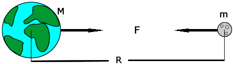
When we usually talk of Gravitation we are bound to think like Newton, where objects are assumed to exerting a force upon each other.
Like imaginary arrows of force in space. But this picture, although good for high school crumbled, with the advent of Einstein’s theory of Relativity.
What is the Space-Time Fabric?
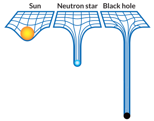
Think of space-time fabric as an actual cloth of fabric. ( An analogy )
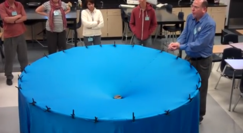
When you place an object on the fabric, the cloth curves. This is exactly what happens in the solar system as well.

The sun with such a huge mass bends the space-time fabric. And the earth and all the planets are kept in orbit by following this curvature that has been made by the sun.
Attributing to the various masses of objects, the way they bend this fabric also varies.
What are Gravitational Waves?
If you drop an object in a medium such as water, they produce ripples that propagate as waves through the medium.

Similarly, Gravitational waves are ripples in space-time fabric produced when you drag heavy objects through space time.
And the nature of these waves is that they don’t require a medium to propagate.
How do you make one?
Everything with mass/energy can create these waves.

Source
Two persons dancing around each other in space too can create gravitational waves. But the waves would be extremely faint.
You need something big and massive accelerating through space-time in order to even detect them.

And orbiting binary stars/black holes are valuable in this retrospect.
How can you detect them?
Let’s turn to the problem to detecting them assuming you do find binary stars/black-holes in the wondrous space to suite your needs.
Well, for starters you cannot use rocks/ rulers to measure them because as the space expands and contracts, so do the rocks. ( the distances will remain same in both the cases )

Here’s where the high school fact that the speed of Light is a constant no matter what plays an important and pivotal role.
If the space expands, the time taken for light to reach from A to B would be longer. And if it contracts, the time taken for it to reach from A to B would be smaller.

PC: PHDComics
By allowing the light waves from the contraction and expansion to interfere with each other, such as done in any interferometry experiment we can detect the expansion or contraction. Voila!
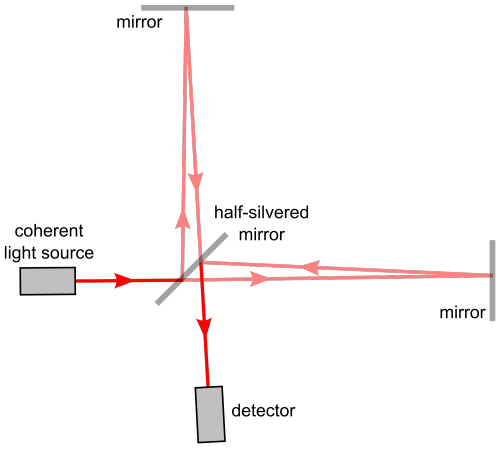
And this is exactly what they did! ( on a macroscopic level ) at LIGO (Laser Interferometer Gravitational-Wave Observatory)
14 September 2015

Two Black Holes with masses of 29 and 36 solar masses merged together some 1.3 Billion light years away.
Two Black Holes colliding is the header animation of the ‘Black Holes are not so Black Series’, in case if you haven’t noticed.

The merger of these two black holes results in the emission of energy equivalent to 3 solar masses as Gravitational Waves.
This signal was seen by both LIGO detectors, in Livingston and Hanford, with a time difference of 7 milliseconds.
And with the measurement of this time difference, physicists have pronounced the existence of Gravitational Waves.
Source
All this is most certainly easily said than done and requires meticulous and extensive research, not to mention highly sensitive instruments.
Had they not have measured this time difference, we might have had to wait for the merger for more massive black holes to collide and maybe even build more sensitive instruments to detect these waves.
And Einstein predicted this a 100 years back!

Mind Blown!
Note: Hope you are able to understand and appreciate the profundity of the discovery done by mankind.
** All animations used here are merely for Educational purposes. If you have any issues, please write to us at : 153armstrong@gmail.com
-
 lizz-blizt reblogged this · 2 years ago
 -
lizz-blizt liked this · 2 years ago
-
 doctorkhanbomb liked this · 4 years ago
 -
 fillejondrette reblogged this · 4 years ago
 -
 currentboat reblogged this · 4 years ago
 -
currentboat liked this · 4 years ago
-
 erza155hasleftthebuilding liked this · 5 years ago
 -
 intheashes reblogged this · 5 years ago
 -
 scientificstoner liked this · 6 years ago
 -
 mer-deuropa reblogged this · 6 years ago
 -
 yellowcrayolacrayon reblogged this · 6 years ago
 -
yellowcrayolacrayon liked this · 6 years ago
-
 passionfruitandvelvet reblogged this · 6 years ago
 -
 growthwitch reblogged this · 6 years ago
 -
 bookbunny9 liked this · 6 years ago
 -
 juliasinikka reblogged this · 7 years ago
 -
 jstevensonart liked this · 7 years ago
 -
 daphnectar liked this · 7 years ago
 -
 charsiu reblogged this · 7 years ago
 -
 yutke reblogged this · 7 years ago
 -
 fofinha liked this · 7 years ago
 -
 nito-onna reblogged this · 7 years ago
 -
 procatboy liked this · 7 years ago
 -
 yyalu reblogged this · 7 years ago
 -
 doctorwormandtheelectricmayhem reblogged this · 7 years ago
 -
 bbxrnxrdx reblogged this · 7 years ago
 -
 nuu-wa reblogged this · 7 years ago
 -
 xeenoo reblogged this · 7 years ago
 -
 deenique reblogged this · 7 years ago
 -
 chocolateiscool liked this · 7 years ago
 -
 manis6 liked this · 7 years ago
 -
 crybabey reblogged this · 7 years ago
 -
 nostraights reblogged this · 7 years ago
 -
nostraights liked this · 7 years ago
-
 double-wind liked this · 7 years ago
 -
 ilaats liked this · 7 years ago
 -
 moved3 liked this · 7 years ago
 -
 bbyfggt liked this · 7 years ago
 -
 rte2 reblogged this · 7 years ago
A reblog of nerdy and quirky stuff that pique my interest.
291 posts