Molecule Of The Day: VX
Molecule of the Day: VX


VX (C11H26NO2PS) is a colourless, odourless, oily liquid under room temperatures. It is a member of the V-series of nerve agents, and is an extremely potent poison - only 0.01 grams of it is needed to kill a person by skin contact. VX was recently implicated in the assassination of Kim Jong-nam, the half-brother of the North Korean leader Kim Jong-un, in Malaysia.
VX is a potent inhibitor of acetylcholinesterase, which breaks down the neurotransmitter acetylcholine into acetic acid and choline. The normal function of the enzyme is to regulate the concentration of acetylcholine within the synaptic cleft, so as to control the frequency of binding of acetylcholine to cholinergic receptors on the postsynaptic cell membrane and hence the transmission of impulses across the synapse.

Consequently, the inhibition of acetylcholinesterase results in a rapid increase in the synaptic concentration of acetylcholine, as the presynaptic knob continues to synthesise it and secrete it into the synaptic cleft. As a result, the cholinergic receptors on the postsynaptic cell membrane are continually stimulated, and a rapid series of action potentials are triggered. This results in muscle spasms and eventual paralysis, leading to death by asphyxiation due to paralysis of the diaphragm.

VX exposure is usually treated using an injection of atropine and pralidoxime. Atropine inhibits certain cholinergic receptors, reducing the binding of acetylcholine to receptors and thus the triggering of action potentials. On the other hand, one end of pralidoxime binds to acetylcholinesterase and the other binds to the phosphate group of VX, which causes the VX molecule to detach from the enzyme together with the pralidoxime molecule (see below). This restores the ability of acetylcholinesterase to hydrolyse acetylcholine, hence reducing its synaptic levels.

VX is synthesised from phosphorus trichloride over multiple steps; first, it is methylated, reacted with ethanol, then transesterified with N,N-diisopropylaminoethanol to produce QL. This is then oxidised with sulfur, and isomerised via heating to produce VX.

More Posts from Contradictiontonature and Others

How the ‘police’ of the cell world deal with 'intruders’ and the 'injured’
The job of policing the microenvironment around our cells is carried out by macrophages. Macrophages are the 'guards’ that patrol most tissues of the body - poised to engulf infections or destroy and repair damaged tissue.
Over the last decade it has been established that macrophages are capable of detecting changes in the microenvironment of human tissues. They can spot pathogen invasion and tissue damage, and mediate inflammatory processes in response, to destroy microbial interlopers and remove and repair damaged tissue. But how do these sentinels of the cell world deal with infection and tissue injury?
Dr Anna Piccinini, an expert in inflammatory signalling pathways in the School of Pharmacy at The University of Nottingham, has discovered that the macrophage’s 'destroy and repair service’ is capable of discriminating between the two distinct threats even deploying a single sensor. As a result, they can orchestrate specific immune responses - passing on information in the form of inflammatory molecules and degrading tissue when they encounter an infection and making and modifying molecular components of the tissue when they detect tissue damage.
Dr Piccinini’s research is published today, Tuesday 30 August 2016, in the academic journal Science Signaling. Her findings could provide future targets for the treatment of diseases with extensive tissue damage such as arthritis or cancer where inflammation plays an increasingly recognized role.
Science Signaling
Macrophage Engulfing Bacteria, Artwork by David Mack

A mighty membrane that twists and turns through the gut is starting the new year with a new classification: the structure, called the mesentery, has been upgraded to an organ.
Scientists have known about the structure, which connects a person’s small and large intestines to the abdominal wall and anchors them in place, according to the Mayo Clinic. However, until now, it was thought of as a number of distinct membranes by most scientists. Interestingly, in one of its earliest descriptions, none other than Leonardo da Vinci identified the membranes as a single structure, according to a recent review.
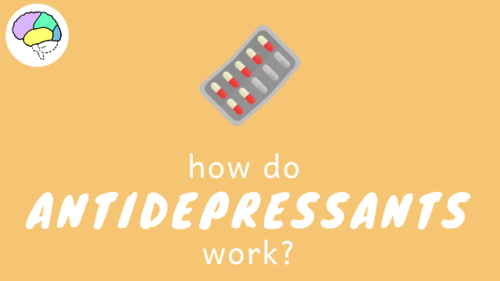

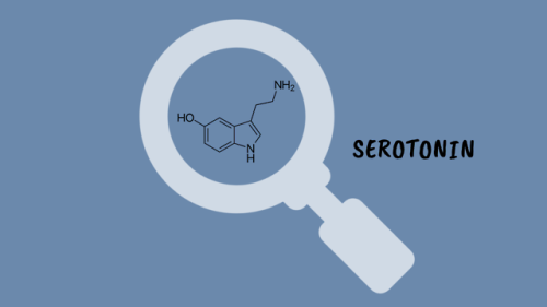
How Do Antidepressants Work? (Video)
Your brain is a network of billions of neurones, all somehow connected to each other. At this very second, millions of impulses are being transmitted through these connections carrying information about what you can see and hear, as well as your emotional state. It’s an incredibly complex system but sometimes things go wrong. Despite extensive research, we are still not certain on the biology that underlies mental illnesses- including depression. However, we have come pretty far in developing effective treatments.

Marrow Christmas and a Happy New Smear!
A very seasonal smear made from red marrow extracted from the iliac crest of a donor’s pelvis prior to transplantation.
Happy Holidays everyone
i♡histo
The image amazingly captures a single moment in time during the development of thousands of red and white blood cells.
Many of the small cells that are visible, like the ones forming the snowman’s carrot nose, do not have a nucleus. These are brand new erythrocytes (red blood cells) that are ready to exit the bone and enter the blood stream.
The other, slightly larger cells that have nuclei, like the snowman’s eyes and his top button, are either precursors to these erythrocytes (they will mature and lose their nucleus) or are precursors to the other blood cells in our body, the leukocytes (white blood cells): lymphocytes, monocytes, neutrophils, eosinophils and basophils.
In addition, the bone marrow is home to the cells that form platelets. These are huge multinucleated cells aptly named megakaryocytes - perhaps the cell at the bottom right.
It is possible to identify each mature cell and its precursor based upon its morphology and staining at higher magnification. High or low levels of these cells can indicate disease or cancers of the blood.
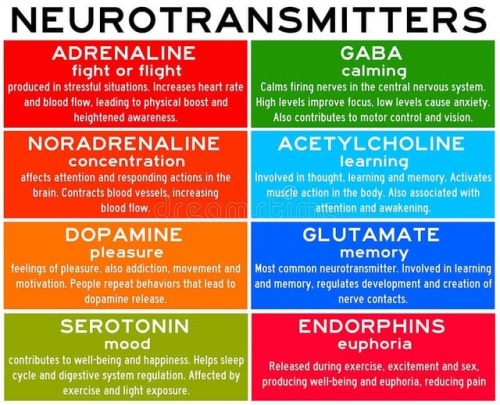
Neurotransmitters are chemicals that help in transmitting signals across a synapse. Different neurotransmitters are associated with different functions. Knowledge about these helps us to treat various neurological conditions by either stimulating or inhibiting these production. #neurology #neuroscience #psychiatry #medicine #medstudynotes #medschool #mbbs #unimed #brain #nervoussystem #physiology #medblog #medblr #medstudent https://www.instagram.com/p/BrM4ocsBqJe/?utm_source=ig_tumblr_share&igshid=12tojib83c32d


Scientists are pretty sure that deep inside the moon, there’s water
While Earth’s surface cracks and spouts fire, the moon’s surface, for as long as we’ve known it, has been quiet.
The youngest sign of volcanic activity scientists have found on the moon’s surface is 18 million years old.
But the traces of that long-ago volcanic activity could help scientists crack an enduring mystery: How much water is on the moon?
A study published Monday in Nature Geoscience suggests it may be more than we thought. Read more (7/24/17)
follow @the-future-now
Are colour-changing octopuses really colourblind?
Cephalopods, including octopuses and squid, have some of the most incredible colour-changing abilities in nature.
They can almost instantly blend in with their surroundings to evade predators or lay in wait, and put on colourful displays to attract mates or dazzle potential prey.
This is impressive enough on its own, but becomes even more amazing when you discover these creatures are in fact colourblind – they only have one type of light receptor in their eyes, meaning they can only see in black and white.
So how do they know what colours to change to at all?
This has puzzled biologists for decades but a father/son team of scientists from the University of California, Berkeley, and Harvard University think the unusual shape of their pupils holds the key, and they can see colour after all.
Cephalopods have wide U-shaped or dumbbell-shaped pupils, which allow light into the lens from many directions.

When light enters the pupils in human eyes it gets focused on one spot, cutting down on blur from the light being split into its constituent colours.
The scientists believe cephalopod eyes work the opposite way – the wide pupils split the light up and then individual colours can be focused on the retina by changing the depth of the eyeball and moving the pupil around.
The price for this is blurry vision, but it does mean they could make out colours in a unique way to any other animals.
Processing colour this way is more computationally intensive than other types of colour vision and likely requires a lot of brainpower, which might explain in part why cephalopods are the most intelligent invertebrates on Earth.

Read the paper
Images: Roy Caldwell, Klaus Stiefel, Alexander Stubbs
Thumpety thump thump thumpety thump thump look at Kinesin go
Myosin, kinesin, and dynein are important proteins governing internal transport. Myosin attached to organelles associates with actin microfilaments to enable the continuous flow of cytoplasm called cytoplasmic streaming.
Kinesins and dynein enable the movement of organelles along microtubules. They attach and move along microtubules. Most kinesins transport organelles from the center towards the periphery of the cell, anterograde transport. Dynein, and a few types of kinesins transport towards the cell center, retrograde transport.
(via thelifeofapremed)

How are elements created in space, stars, and in laboratories? The latest edition of #PeriodicGraphics in C&EN takes a look! http://bit.ly/2UXWoPD http://bit.ly/2YbuBNE
-
 godverdomme-toch reblogged this · 3 years ago
 -
 kristallijne-diamorphine reblogged this · 4 years ago
 -
 pleasurehunter2000 liked this · 4 years ago
 -
man-in-the-desert liked this · 5 years ago
-
 vidavelha reblogged this · 5 years ago
 -
 010100 liked this · 6 years ago
 -
 professional-dragonsandmemes liked this · 6 years ago
 -
 believe3141984 liked this · 6 years ago
 -
 moo-stew liked this · 6 years ago
 -
 tiltingatwindmills90 reblogged this · 6 years ago
 -
tiltingatwindmills90 liked this · 6 years ago
-
 turn0nthedarkness reblogged this · 6 years ago
 -
turn0nthedarkness liked this · 6 years ago
-
 little-ursa liked this · 6 years ago
 -
salvamarmolejo liked this · 6 years ago
-
 fireofdevoti0n liked this · 6 years ago
 -
 hematwodd-blog liked this · 6 years ago
 -
 iguessimsorryjenni reblogged this · 6 years ago
 -
floral-mask liked this · 7 years ago
-
 sssrst-blog liked this · 7 years ago
 -
 purplebee13 liked this · 7 years ago
 -
 like-nxrthernstxrs reblogged this · 7 years ago
 -
perpendicularthoughts reblogged this · 7 years ago
-
 mxviatrix reblogged this · 7 years ago
 -
 dilly2dally reblogged this · 7 years ago
 -
dilly2dally liked this · 7 years ago
-
dendrobateno liked this · 7 years ago
-
 belikegregory liked this · 7 years ago
A pharmacist and a little science sideblog. "Knowledge belongs to humanity, and is the torch which illuminates the world." - Louis Pasteur
215 posts